Foot Bone Anatomy Vector Illustration 539973 Vector Art at Vecteezy

Foot Bone Anatomy Vector Illustration 539973 Vector Art at Vecteezy
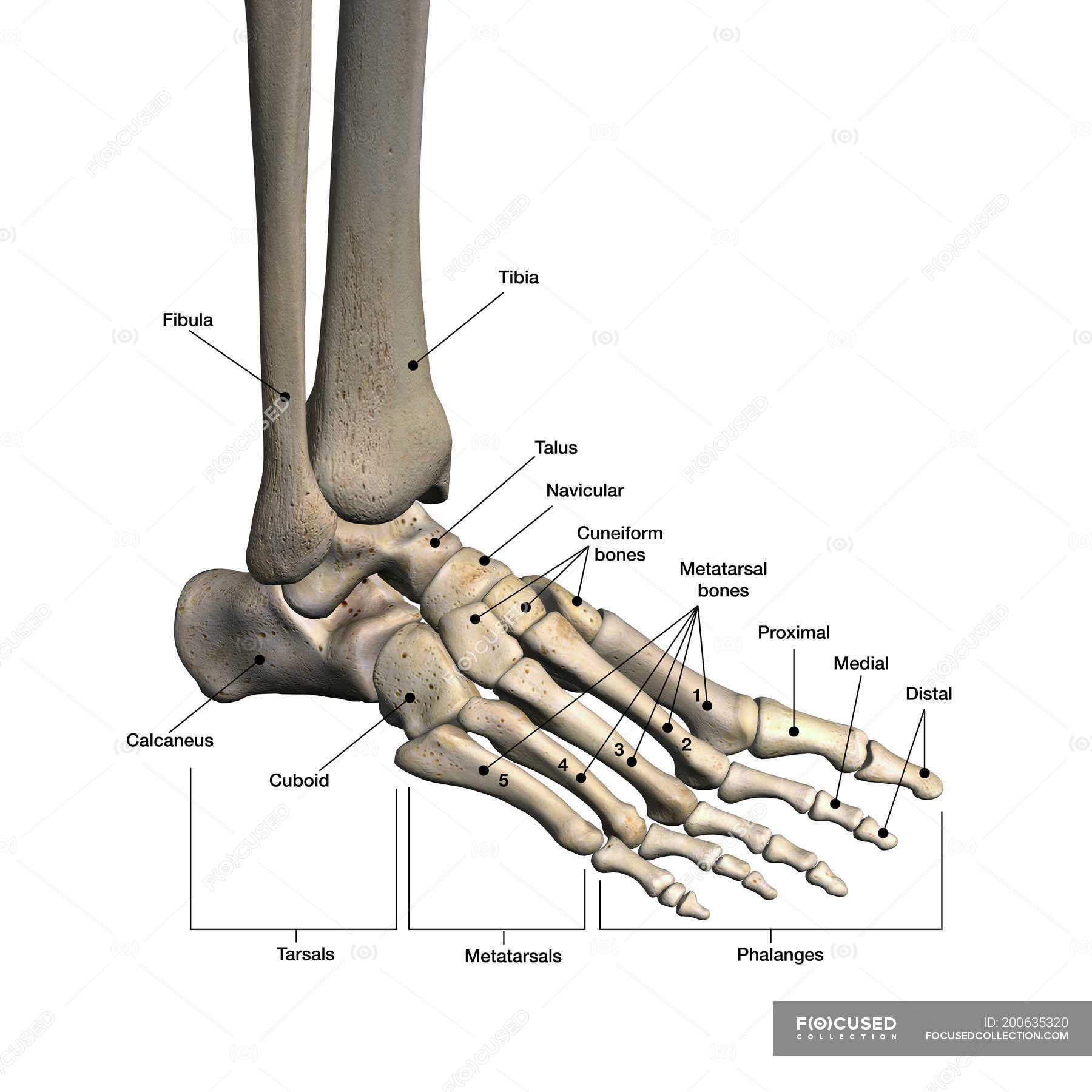
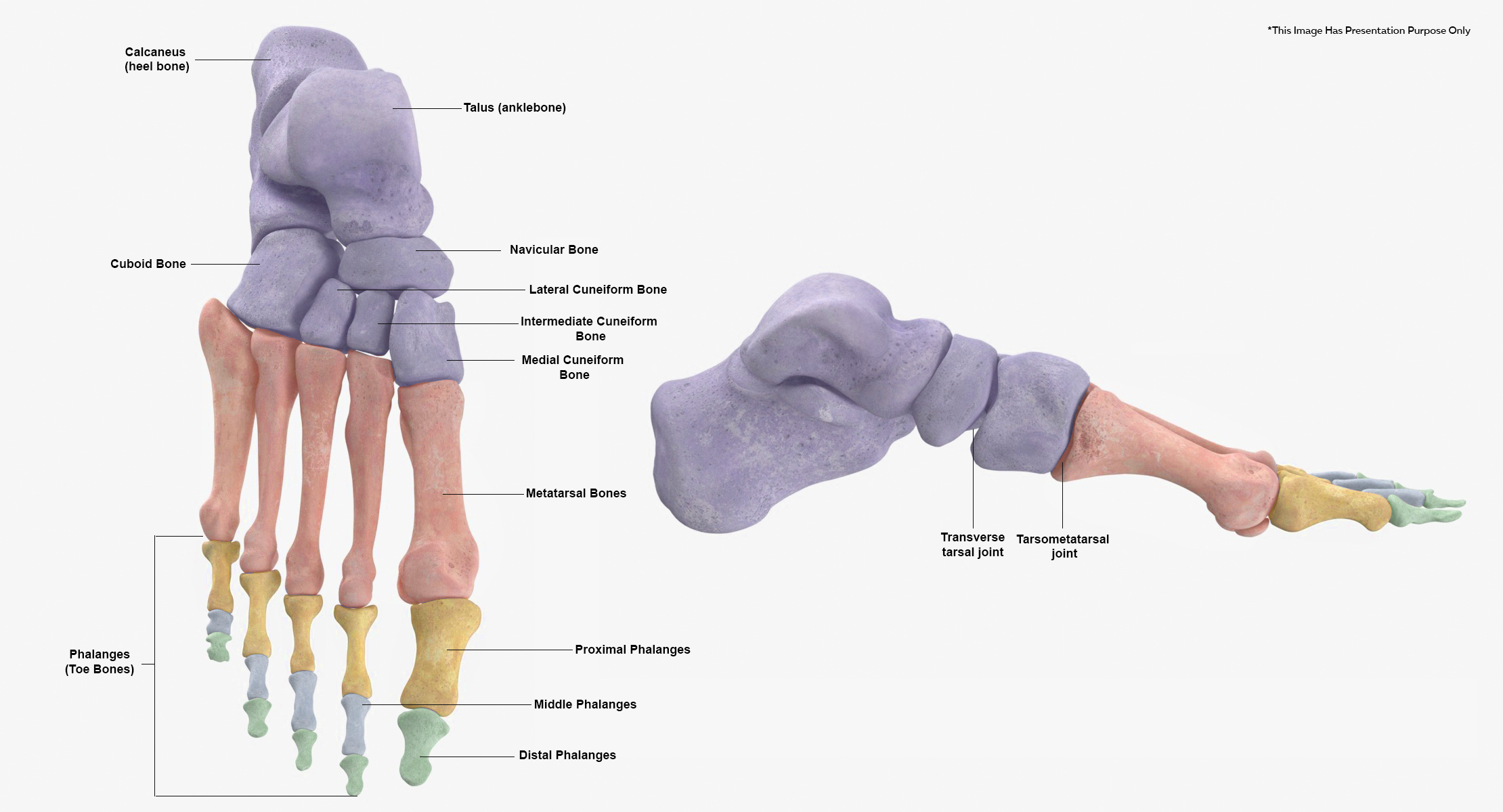
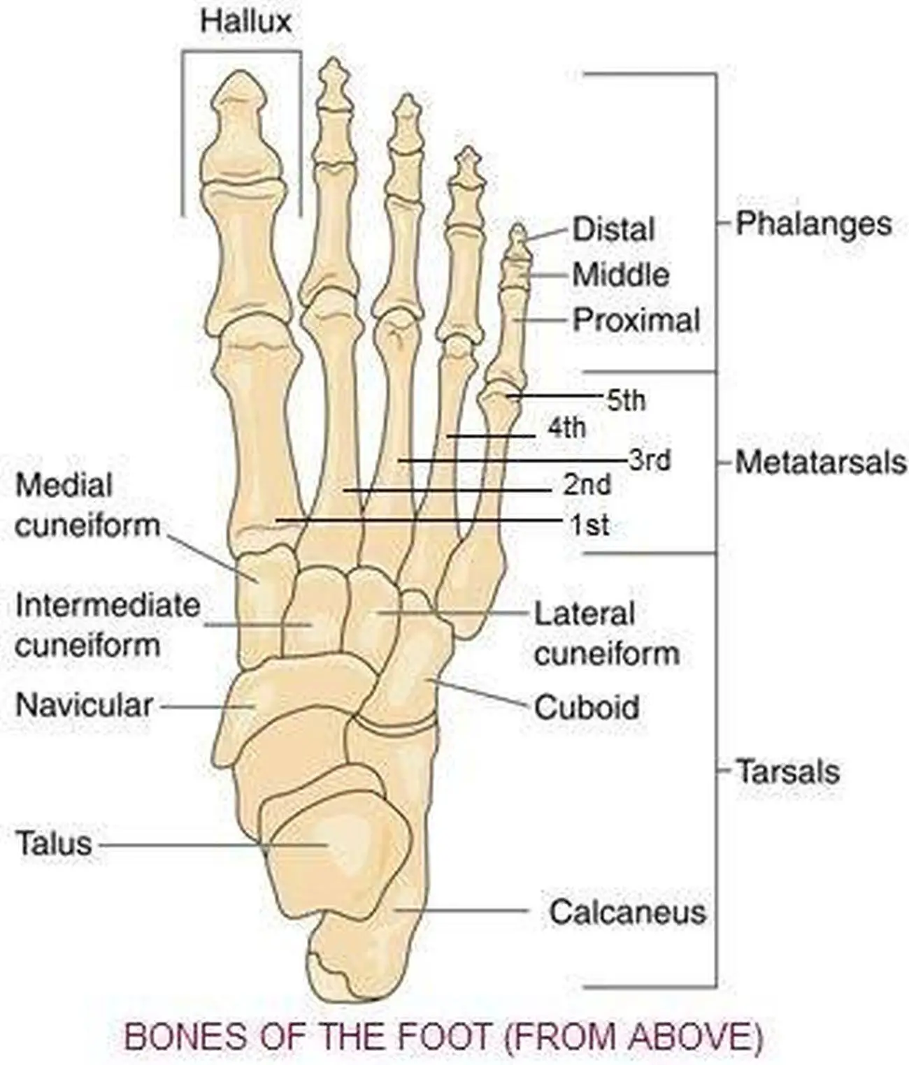
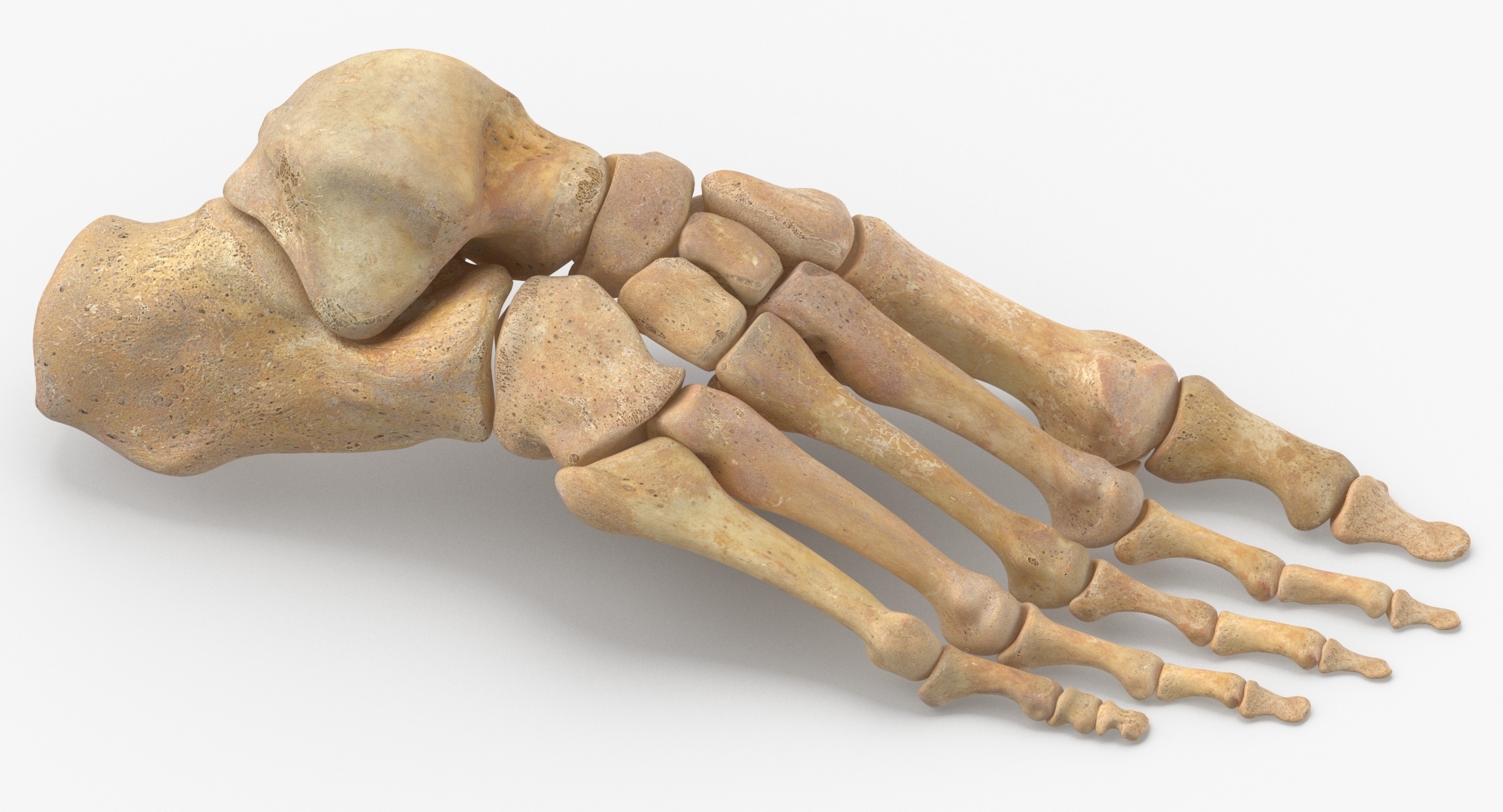
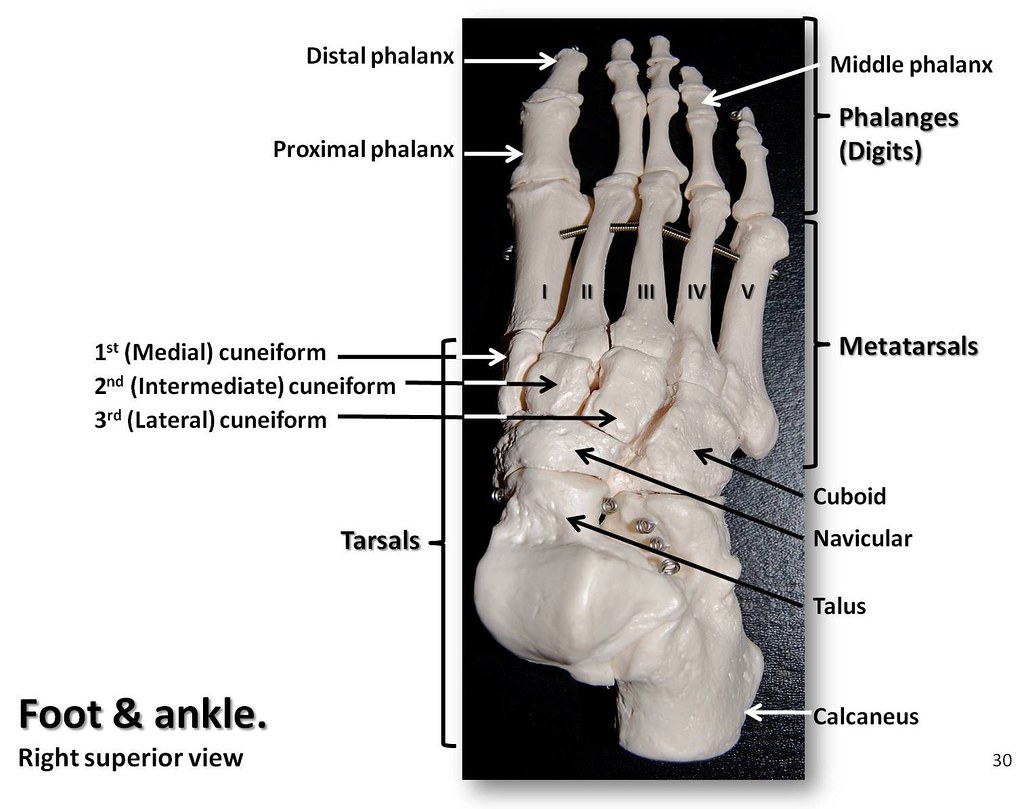
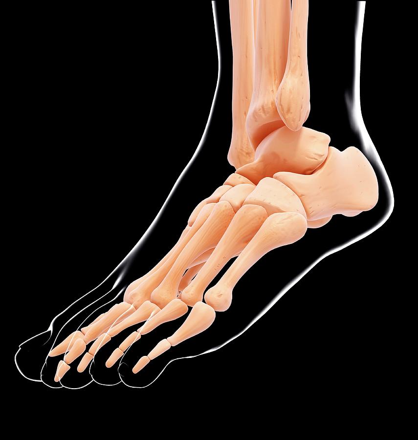
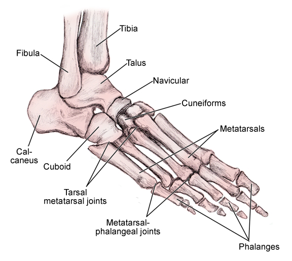
The bones of the foot provide mechanical support for the soft tissues; helping the foot withstand the weight of the body whilst standing and in motion.. They can be divided into three groups: Tarsals - a set of seven irregularly shaped bones.They are situated proximally in the foot in the ankle area. Metatarsals - connect the phalanges to the tarsals.
Bones of human foot with labels on white background — phalanx, fibula
kool99/Getty Images In the foot, there are: 26 bones 33 joints more than 100 muscles, tendons, and ligaments Bones of the foot The bones in the foot make up nearly 25% of the total.

Right Foot Bones
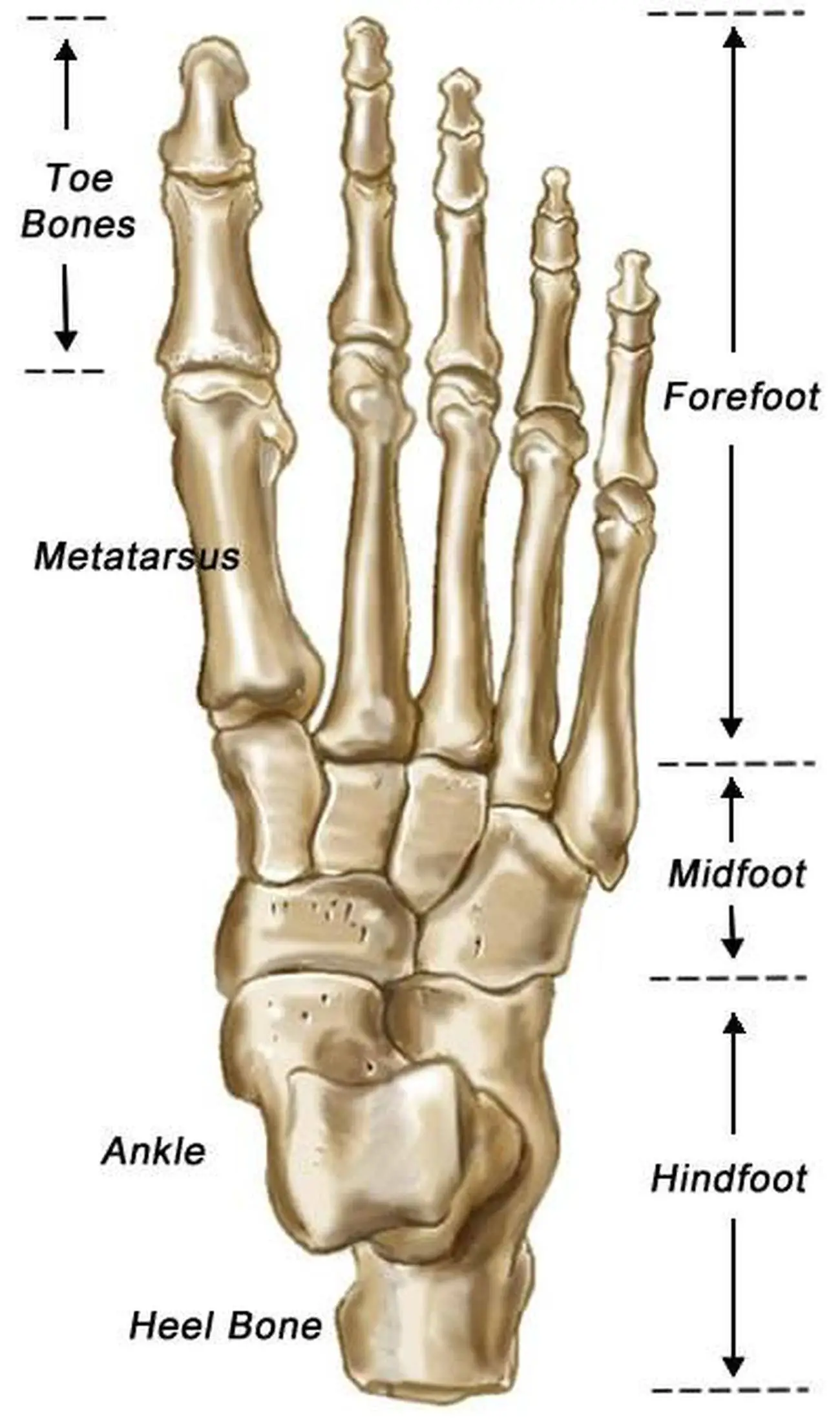
Last updated 2 Nov 2018 The anatomy of the foot The foot contains a lot of moving parts - 26 bones, 33 joints and over 100 ligaments. The foot is divided into three sections - the forefoot, the midfoot and the hindfoot. The forefoot
3D Human Foot Tarsals and Metatarsal Bones Collection Yellow 12
Hand on foot as suffer from inflammation feet problem of Sever's Disease or calcaneal apophysitis. human foot bones stock pictures, royalty-free photos & images Heel Pain or plantar fasciitis concept.
Pictures Of Bones Of The Feet
Realistic foot bones anatomy set with isolated side views of human footstep skeleton on blank background vector illustration Plantar fasciitis. Plantar fascia inflammation or tearing. Disorder of connective ligament which supports the arch of the foot. Plantar heel pain syndrome. Flat vector illustration
Human foot bones anatomy 3D model TurboSquid 1558150
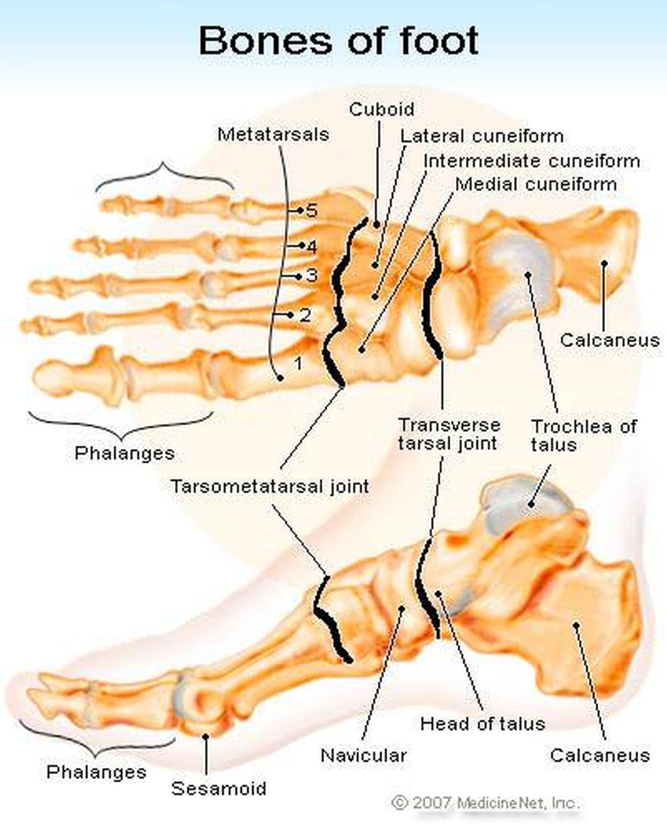
Image credit: Stephen Kelly, 2019 Tarsal bones The tarsal bones are a group of seven bones that make up the rear section of the foot. Tarsal bones include: The talus, or ankle bone: The.
Bones of the foot and ankle, superior view with labels Appendicular
2,836 Human Foot Bones Stock Photos, High-Res Pictures, and Images - Getty Images Boards Sign in Browse Creative Images Creative Images Browse millions of royalty-free images and photos, available in a variety of formats and styles, including exclusive visuals you won't find anywhere else. See all creative images Trending Image Searches

Foot Fracture
The navicular bone is found on the inner side of the foot. The navicular articulates with five of the other tarsal bones - at the top with the talus, talonavicular joint, laterally (outer side) with the cuboid, cubonavicular joint, and at the bottom it articulates with the three cuneiform bones. In around 10% of the population, a small extra piece of bone develops next to the navicular which.
Pictures Of Bones Of The Feet
The foot (plural feet) is an anatomical structure found in many vertebrates. It is the terminal portion of a limb which bears weight and allows locomotion. In many animals with feet, the foot is a separate organ at the terminal part of the leg made up of one or more segments or bones, generally including claws or nails.
Left human foot bones Buy Royalty Free 3D model by Catherine Sulzmann
The ankle joint The superior tibiofibular joint (joint near the knee that holds the tibia and fibula together) The inferior tibiofibular joint (the lower joint at the ankle that holds the tibia and fibula together)

The Bones Of The Foot Digital Art by
Browse 4,222 foot bones photos and images available, or search for human foot bones to find more great photos and pictures. Browse Getty Images' premium collection of high-quality, authentic Foot Bones stock photos, royalty-free images, and pictures. Foot Bones stock photos are available in a variety of sizes and formats to fit your needs.

Ankle and Foot Pain Massage Therapy Connections
Foot: The end of the leg on which a person normally stands and walks. The foot is an extremely complex anatomic structure made up of 26 bones and 33 joints that must work together with 19 muscles and 107 ligaments to execute highly precise movements. At the same time the foot must be strong to support more than 100,000 pounds of pressure for.
Pictures Of Bones Of The Feet
The foot contains many bones, muscles, tendons, and other structures. Learn about the anatomy of parts of the foot and common problems that can occur.. X-ray: This standard imaging test uses low-level radiation to take pictures. It can pick up conditions like bone fractures, dislocations, or arthritis damage. Computed tomography (CT).
Human Foot Bones Photograph by Pixologicstudio/science Photo Library
foot, in anatomy, terminal part of the leg of a land vertebrate, on which the creature stands. In most two-footed and many four-footed animals, the foot consists of all structures below the ankle joint: heel, arch, digits, and contained bones such as tarsals, metatarsals, and phalanges; in mammals that walk on their toes and in hoofed mammals.
_en.jpg)
Bones Of The Foot Economics Books
Human body Skeletal System Bones of foot Bones of foot The 26 bones of the foot consist of eight distinct types, including the tarsals, metatarsals, phalanges, cuneiforms, talus,.
foot bones 4 CoreWalking
Cuboid Medial cuneiform Intermediate cuneiform Lateral cuneiform Some people may be born with an extra navicular bone ( accessory navicular) beside the regular navicular bone, on the inside of the foot. This is a normal anatomical variation seen in around 2.5% of the entire population of the US. Metatarsal Bones
