Human lungs infographic Education Illustrations Creative Market
Respiratory System Anatomy and Physiology Nurseslabs
The lungs are a major organ that is part of the respiratory system, taking in fresh air and getting rid of old, stale air. This mechanism of breathing also helps to allow you to talk. By taking in fresh air, the lungs are able to help oxygenate blood to be carried around your body. This is done by inhaling the air and bringing it in toward the.
Lung Info Canadian Lung Association
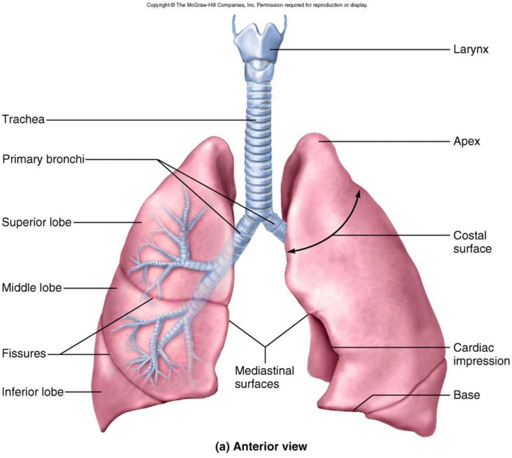
Surfaces. There are three lung surfaces, each corresponding to an area of the thorax. The mediastinal surface of the lung faces the lateral aspect of the middle mediastinum. The lung hilum (where structures enter and leave the lung) is located on this surface.. The base of the lung is formed by the diaphragmatic surface.It rests on the dome of the diaphragm, and has a concave shape.
Lung Diagram Labeled EdrawMax Template
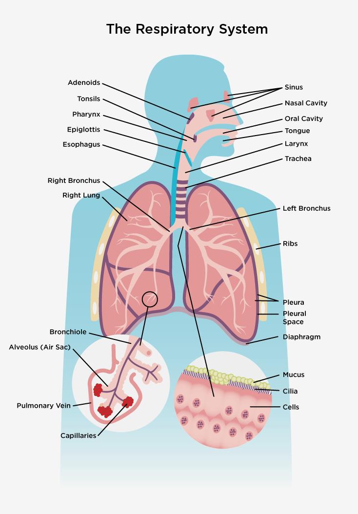
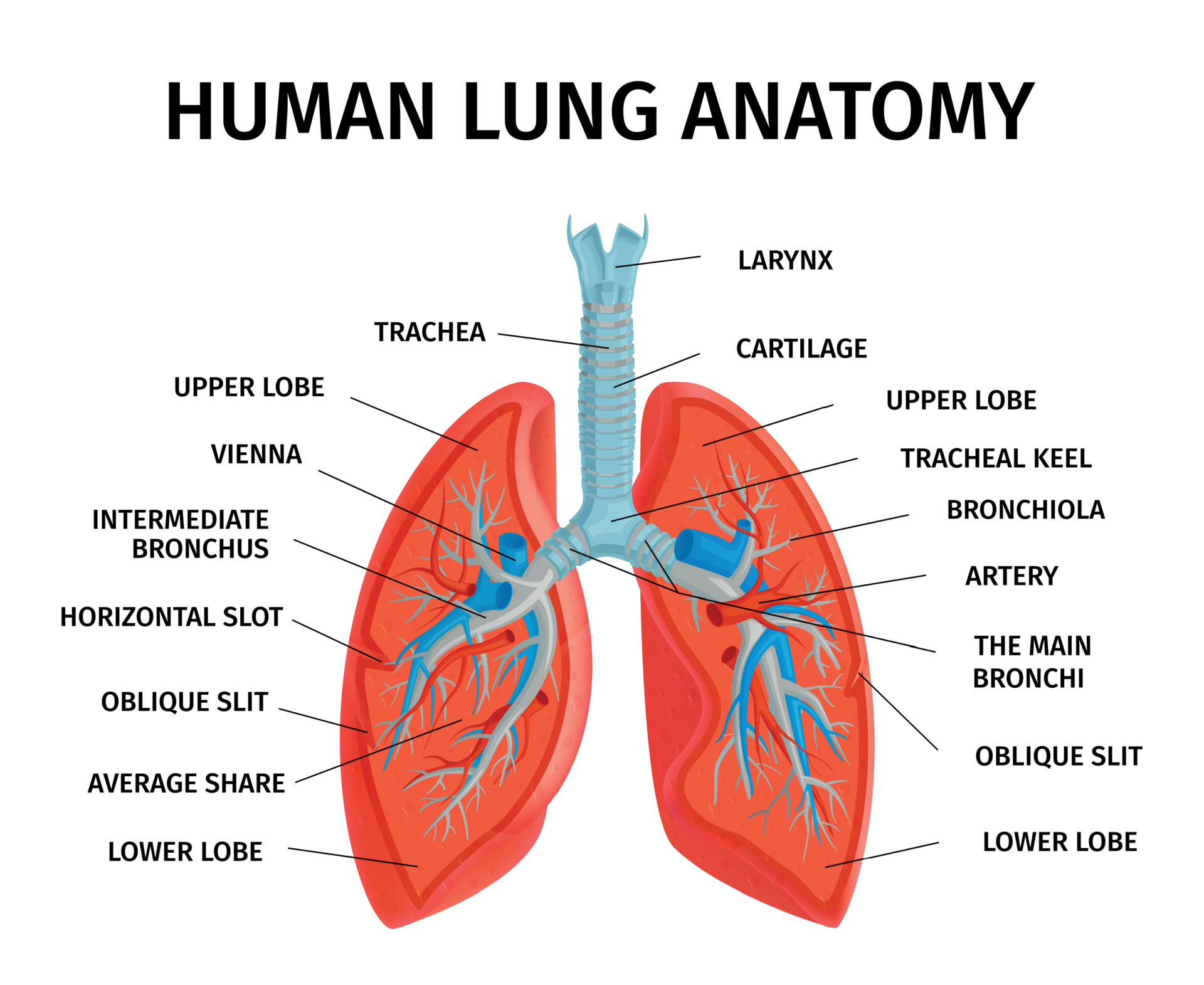
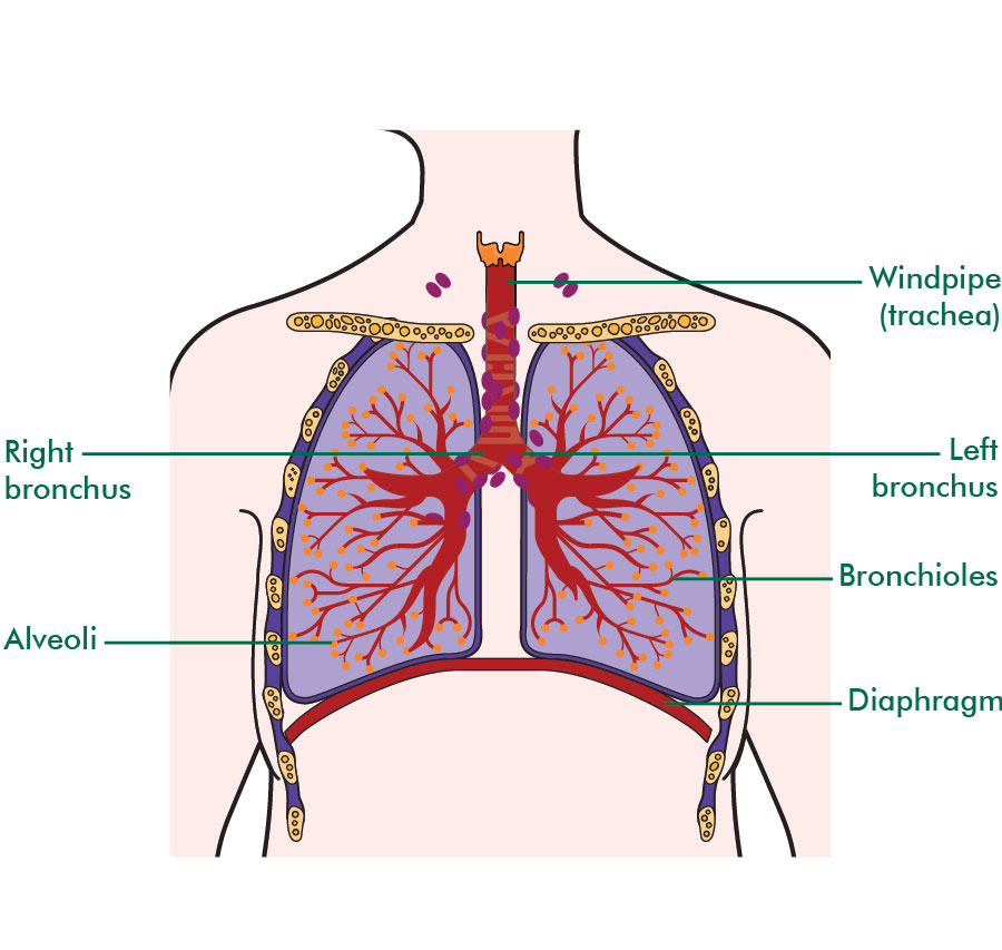
The lungs are respiratory organs - we use them to breathe. We breathe in order to get oxygen (O 2) and to get rid of carbon dioxide (CO 2 ). We breathe in through the nose or mouth. The air then goes through the larynx and the trachea (also called the windpipe) and into the lungs. We breathe by using the diaphragm, a muscular membrane under.

Lung Structure BioNinja
View Original Image at Full Size. Labeled diagram of the lungs/respiratory system. Image 37789 is a 1125 by 1408 pixel WebP Uploaded: Jan10 14. Last Modified: 2014-01-10 12:15:34
The structure of a lung with labeled parts. Biology vector illustration vector de Stock Adobe
Damaged lungs are darker gray and can have black spots in them. Your triangularly shaped right and left lungs look a little bit like the ears of an elephant. A typical lung in a human adult lung weighs about 2.2 pounds and is a little longer than 9 inches when you're breathing normally, and about 10.5 inches when your lungs are completely.
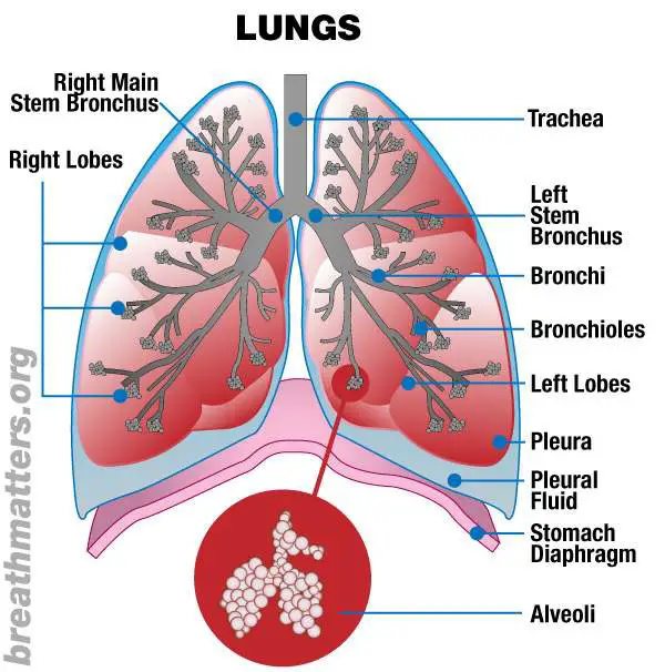
Diagram of lungs
Lungs are a pair of respiratory organs situated in a thoracic cavity. Right and left lung are separated by the mediastinum. Texture -- Spongy. Color - Young - brown. Adults -- mottled black due to deposition of carbon particles. Weight-. Right lung - 600 gms. Left lung - 550 gms.
The lungs Lung cancer Macmillan Cancer Support
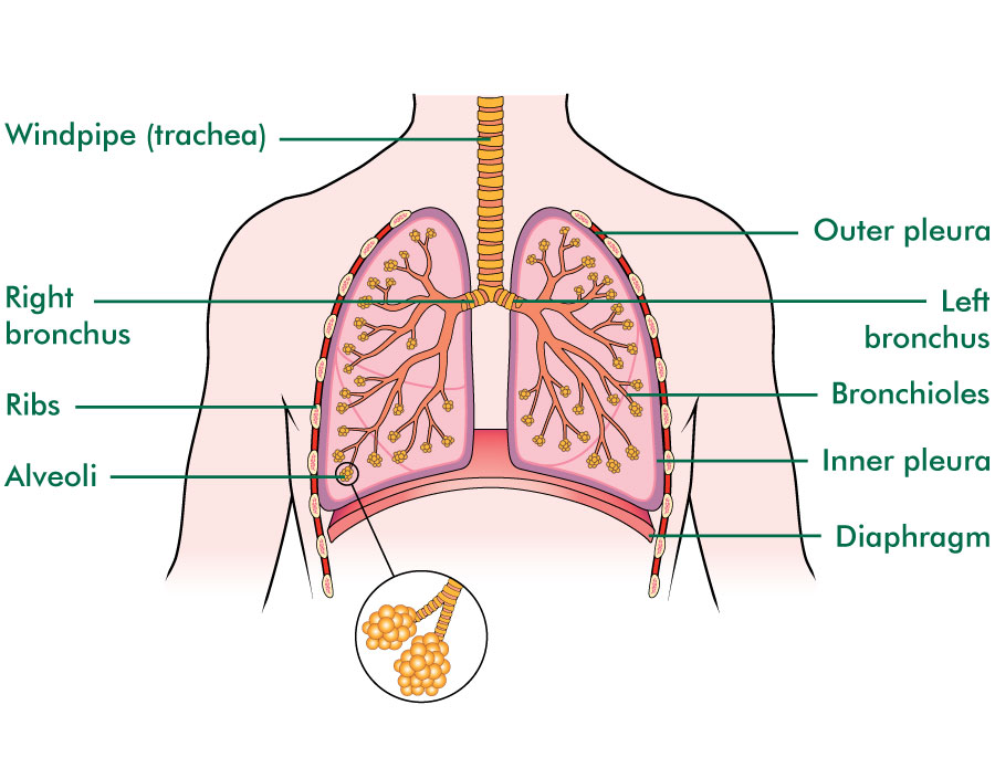
The lungs begin at the bottom of your trachea (windpipe). The trachea is a tube that carries the air in and out of your lungs. Each lung has a tube called a bronchus that connects to the trachea.
CrossFit Anatomy of the Lungs
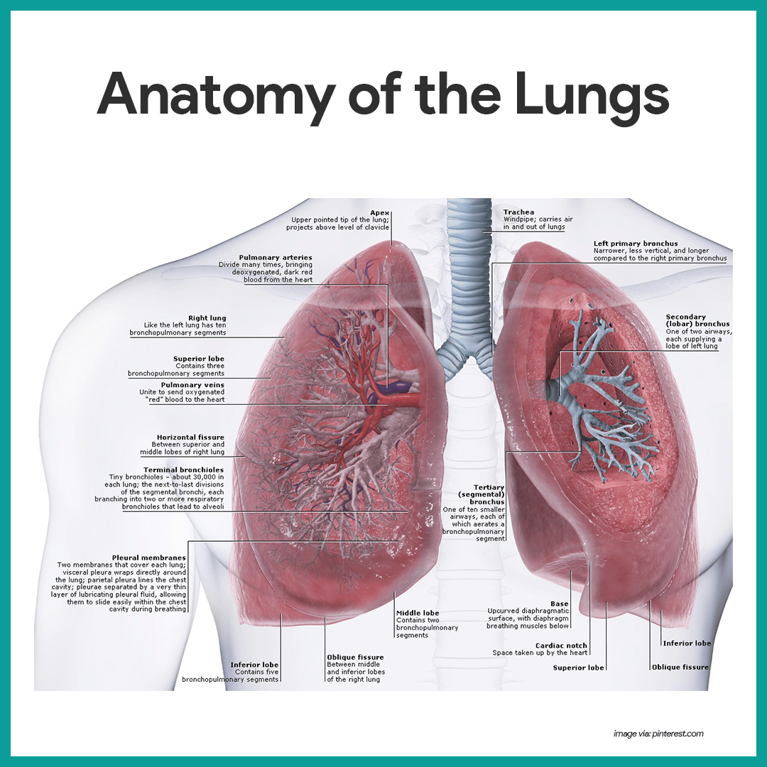
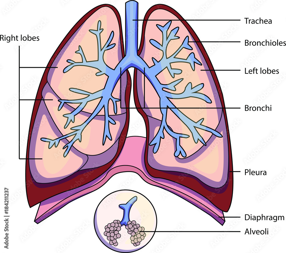
bronchial tree: the system of airways within the lungs, which bring air from the trachea to the lung's tiny air sacs (alveoli). cardiac notch: the indentation in the left lung that provides room for the heart. diaphragm: a muscular membrane under the lungs. larynx: a muscular structure at the top of the trachea, containing the vocal cords.
Pneumothorax Causes, Signs, Symptoms, Treatment
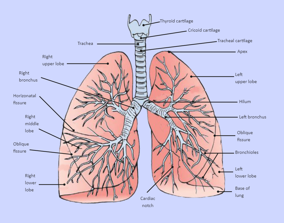
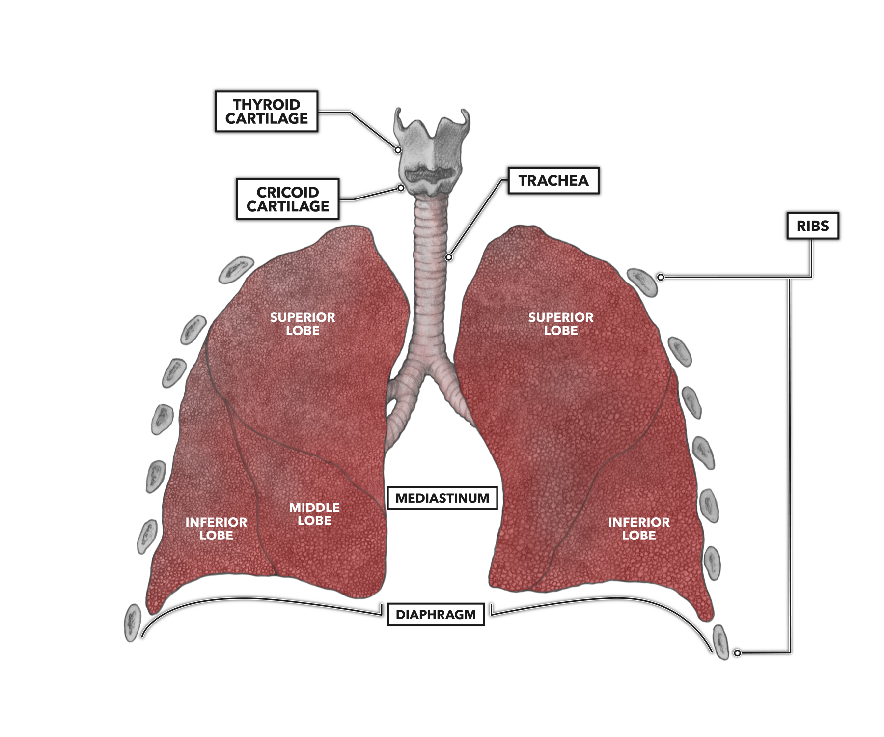
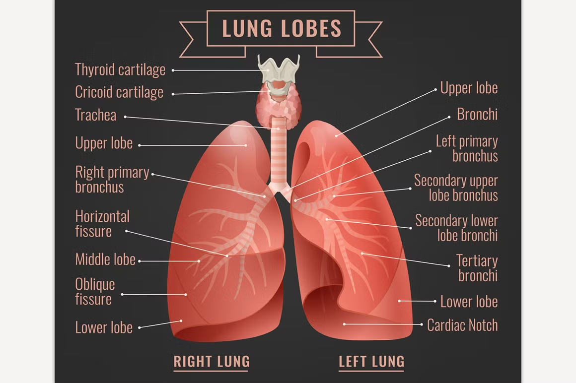
Root of lung. Lingula of left lung. Superior lobe (upper lobe) Middle lobe (only on right) Inferior lobe (lower lobe) Oblique fissure. Horizontal fissure of right lung. 144.30 Main bronchus. 152.23 Pulmonary ligament.
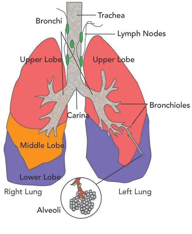
Diagram of lungs
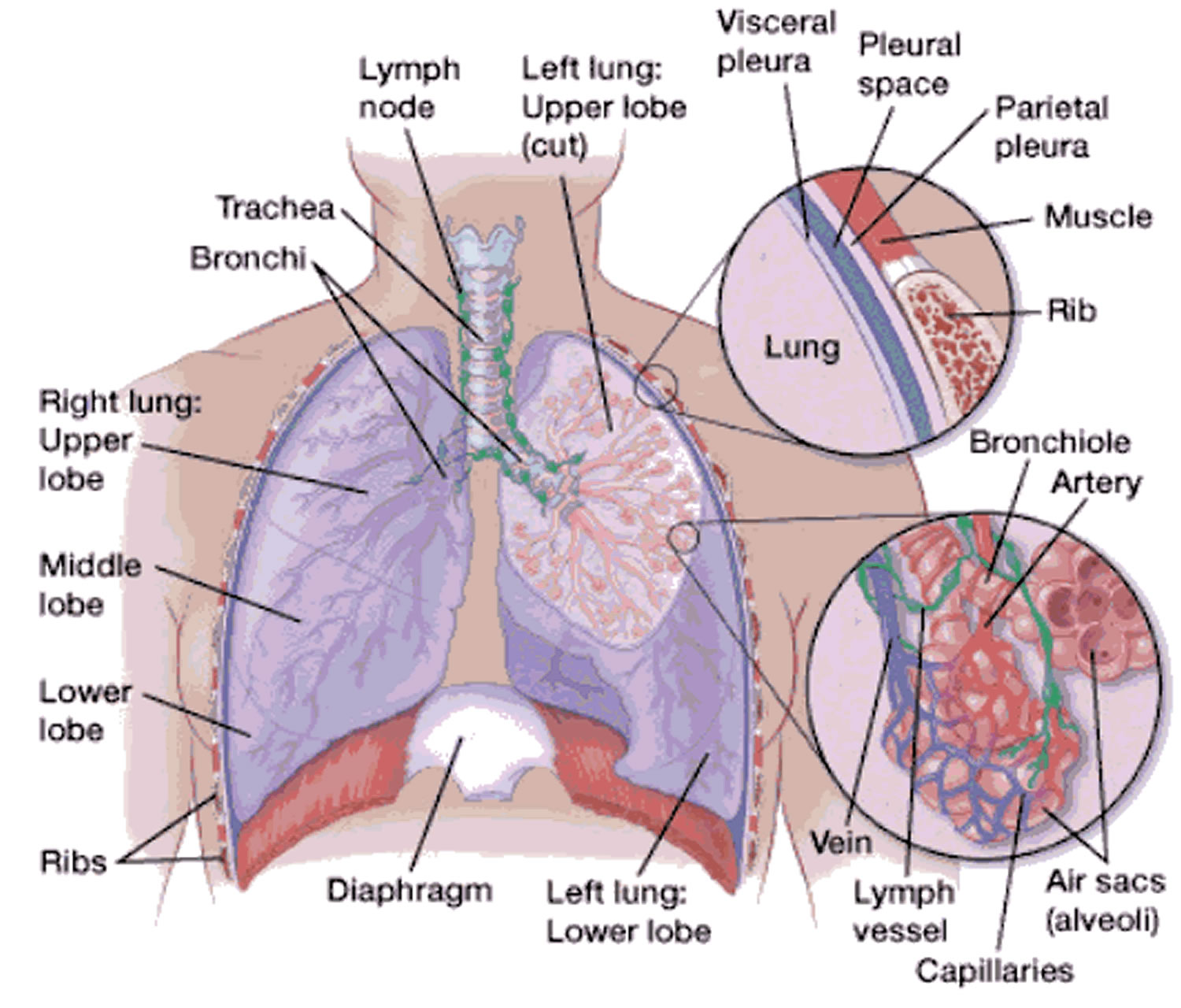
The costal surface of the lung borders the ribs. The mediastinal surface faces the midline. Figure 22.2.1 Gross Anatomy of the Lungs. Each lung is composed of smaller units called lobes. Fissures separate these lobes from each other. The right lung consists of three lobes: the superior, middle, and inferior lobes.
Human Lung Anatomy Diagram 4958464 Vector Art at Vecteezy
The main function of the lungs is to exchange oxygen and carbon dioxide in the body. When a person breathes in, oxygen travels down the back of the throat and into the trachea, the windpipe. The.
Anatomy of LUNGS and MCQs for NEET, GPAT,Nursing Exams Gpatindia Pharmacy Jobs, Admissions
Functionally, the lung is divided into a series of bronchopulmonary segments. The bronchopulmonary segments are the largest subdivision of a lobe. They are separated from adjacent segments by connective tissue septa and are also surgically resectable. They are 10 bronchopulmonary segments in the left lung and 8-10 in the left lung [9].
How do we breathe? (Lungs and Pleura) Interactive Biology, with Leslie Samuel
Lung anatomy can get quite complicated extremely quickly. Ease into the topic and cement your knowledge using Kenhub's respiratory system quizzes and labeled diagrams. Regulation of breathing. The breathing cycle is controlled by the respiratory centre located inside the medulla oblongata and the pons of the brain stem. Three major collections.
labelled lungs diagram
Given below is a labeled diagram of the human lungs followed by a brief account of the different parts of the lungs and their functions. Each lung is enclosed inside a sac called pleura, which is a double-membrane structure formed by a smooth membrane called serous membrane. The outer membrane of this structure is called parietal pleura and is.
Human lungs infographic Education Illustrations Creative Market
The respiratory system aids the body in the exchange of gases between the air and blood, and between the blood and the body's billions of cells. It includes air passages, pulmonary vessels, the.

Respiratory System Anatomy
Lungs-lobes: diagrams detailing the various lung lobes (lower lobe, middle-right lobe, left lingula) separated by the pulmonary fissures.. 125 pulmonary anatomical structures were labeled. Those labels are grouped into subcategories, you can hide or show them on the "Anatomical parts" tab: Trachea; Bronchial tree; Alveolus;
