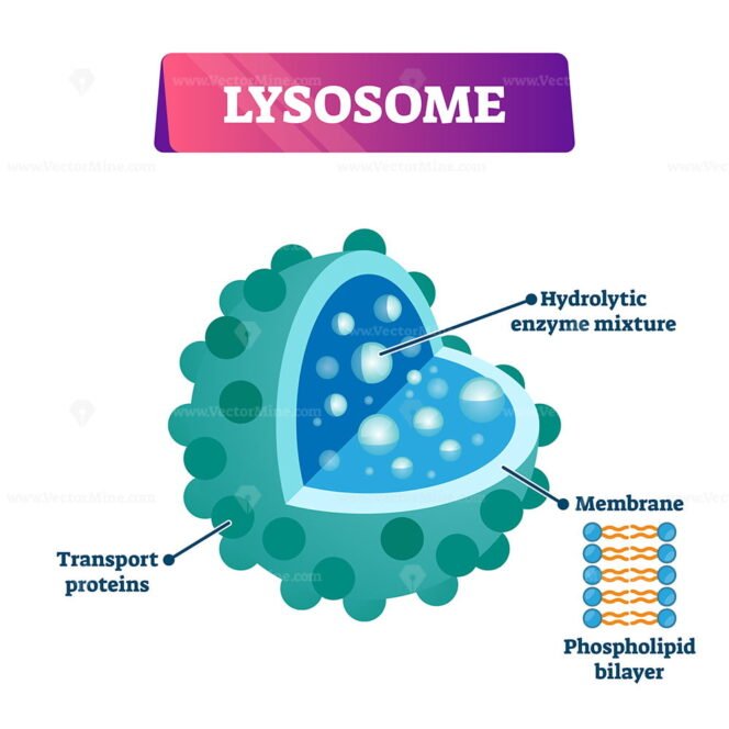
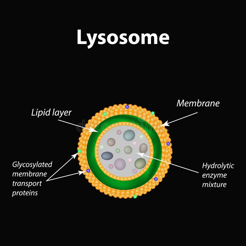
Structure lysosomes infographics Royalty Free Vector Image
Lysosome the cell’s recycling center definition, structure, function, and biology
1) The lysosome is contained by a phospholipid membrane. Why is this membrane important? What would be the effect on the cell if the membrane was broken down? 2) Research at least two types of.

Structure lysosomes infographics Royalty Free Vector Image
By Bert Markgraf Lysosomes are small cell organelles in nucleus-bearing or eukaryotic cells. They are located in the cytosol of the cells, floating freely within the cells outside the nucleus. They have a simple structure made up of an outer lysosomal membrane surrounding an acidic interior fluid.
Marin Science Seminar All About Lysosomes
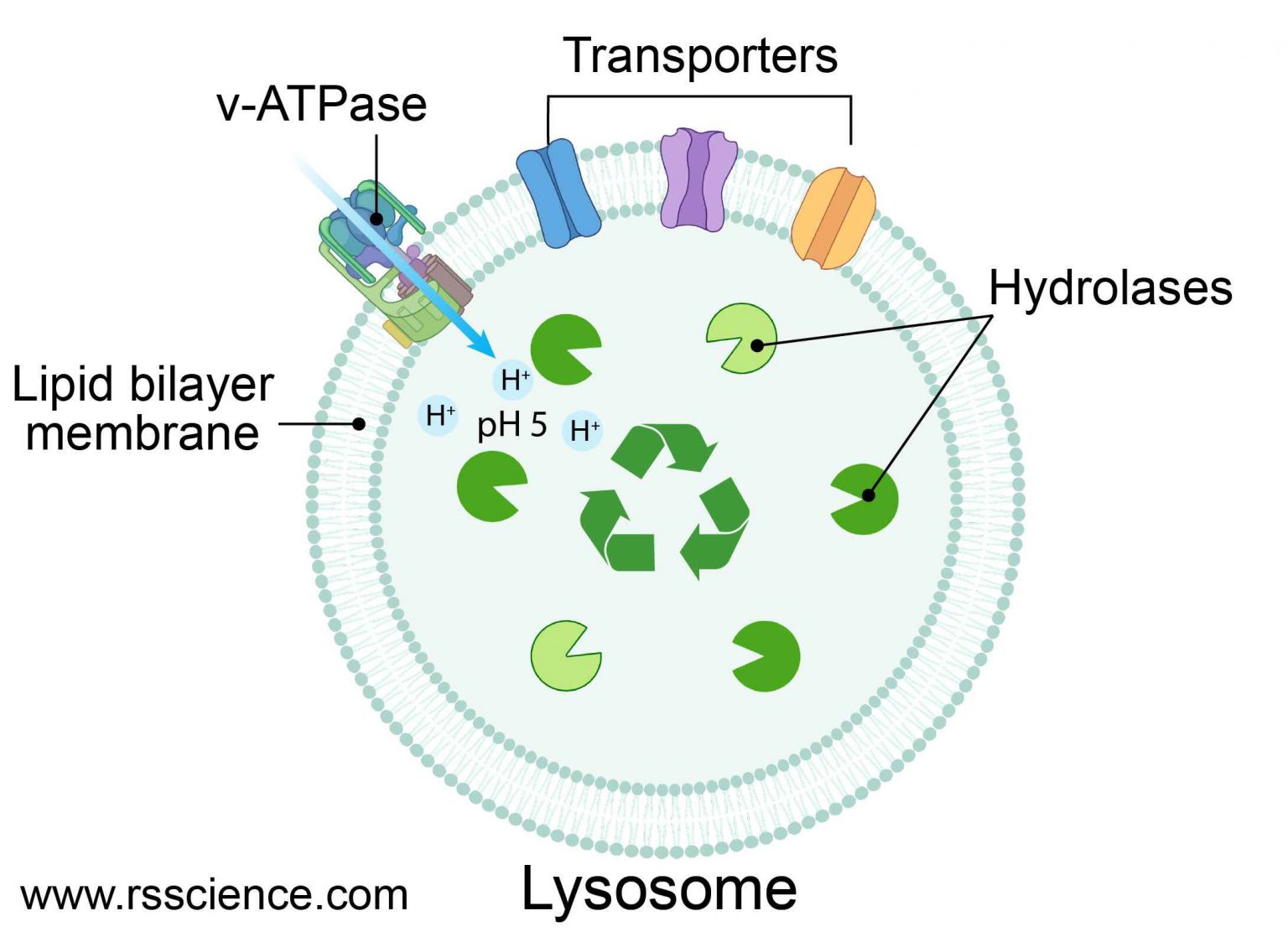
Electron micrograph of lysosomes and mitochondria in a mammalian cell. Lysosomes are indicated by arrows. (Visuals Unlimited/K. G. Murti.) Go to: Lysosomal Acid Hydrolases Lysosomes contain about 50 different degradative enzymes that can hydrolyze proteins, DNA, RNA, polysaccharides, and lipids.
Lysosome cell organelle vector illustration labeled cross section diagram VectorMine
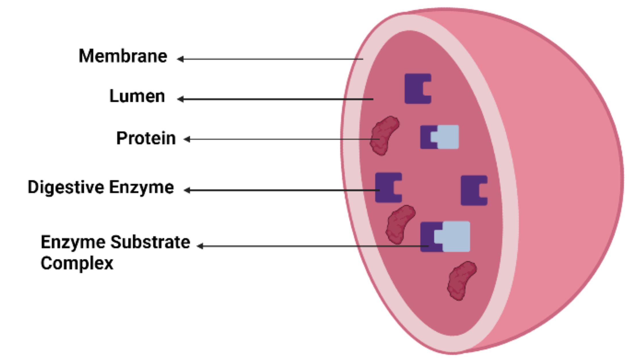
A lysosome ( / ˈlaɪsəˌsoʊm /) is a membrane-bound organelle found in many animal cells. [1] They are spherical vesicles that contain hydrolytic enzymes that digest many kinds of biomolecules. A lysosome has a specific composition, of both its membrane proteins and its lumenal proteins.

General structure and properties of lysosomes. Lysosome lipid bilayer... Download Scientific
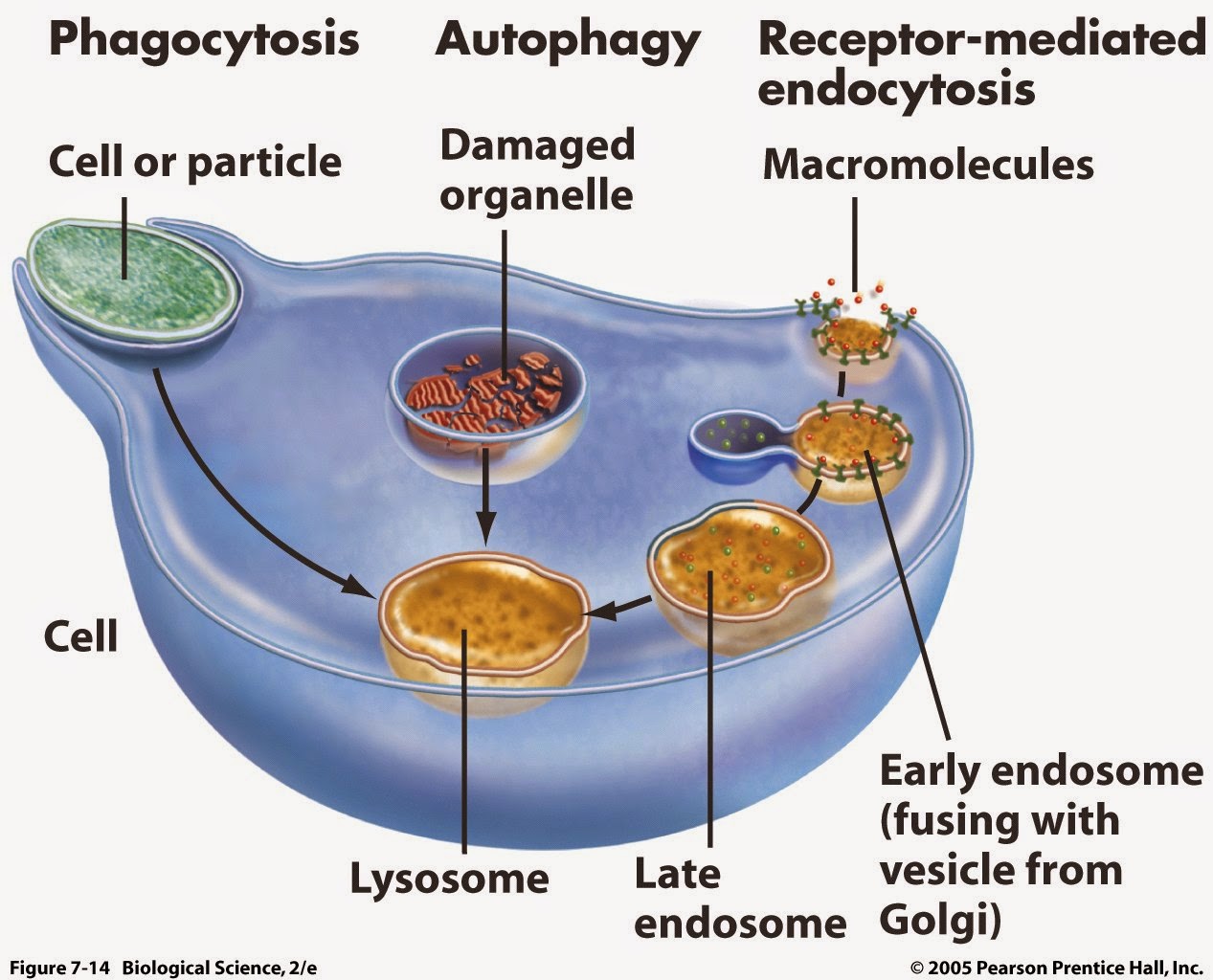
The lysosomes show polymorphism in different cell types. There are two basic types of lysosomes. Golgi complex buds off primary lysosomes containing hydrolytic enzymes. The vacuole or phagosome arising by endocytosis associates and fuses with primary lysosome to form secondary lysosome (Fig. 2.61).
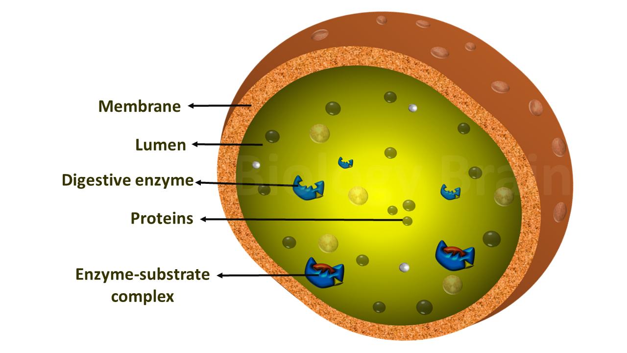
Diagram of Lysosomes and Types Biology Brain
Vesicles and Vacuoles. Vesicles and vacuoles are membrane-bound sacs that function in storage and transport. Vacuoles are somewhat larger than vesicles, and the membrane of a vacuole does not fuse with the membranes of other cellular components. Vesicles can fuse with other membranes within the cell system ( Figure 5.11.1 5.11. 1 ).
Plant Cell Lysosome Is What Are Lysosomes and How Are They Formed? / Cell membrane is the
Key points: All cells have a cell membrane that separates the inside and the outside of the cell, and controls what goes in and comes out. The cell membrane surrounds a cell's cytoplasm, which is a jelly-like substance containing the cell's parts. Cells contain parts called organelles. Each organelle carries out a specific function in the cell.

What is a Lysosome? (with pictures)
Lysosomal biology and function: modern view of cellular debris bin is a review article that summarizes the recent advances in understanding the roles of lysosomes in various cellular processes, such as metabolism, autophagy, immunity and aging. The article also discusses the implications of lysosomal dysfunction in human diseases and potential therapeutic strategies. The article provides a.
The Structure of Lysosomes. Infographics. Vector Illustration Stock Vector Illustration of
Fig 1 - Diagram of the endomembrane system Function The hydrolytic enzymes contained within the lysosome allow foreign particles to be destroyed. Lysosomes play an important role in phagocytosis. When macrophages phagocytose foreign particles, they contain them within a phagosome.

STRUCTURE AND FUNCTIONS OF LYSOSOMES
The endomembrane system ( endo - = "within") is a group of membranes and organelles in eukaryotic cells that works together to modify, package, and transport lipids and proteins.
Lysosomes are practically present in all cells except(A) WBC(B) RBC(C) Epithelial cells(D
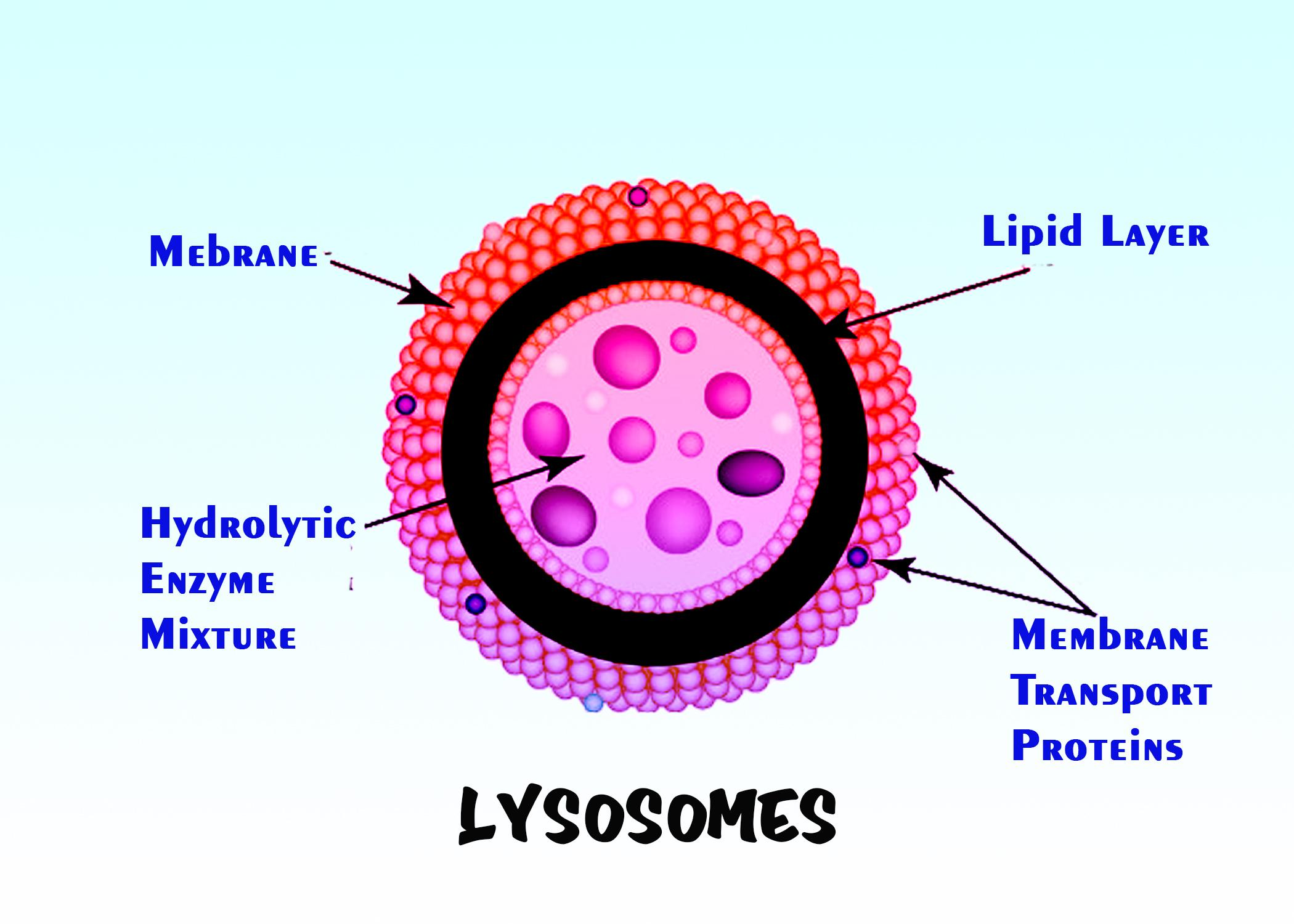
Home Cell Biology Lysosomes: Definition, Structure, Functions Animesh Sahoo November 12, 2021 Lysosomes are tiny vascular membrane-bound vesicles involved in intracellular digestion. They contain a variety of hydrolytic enzymes that remain active under acidic conditions. Greek words " Lysis = digestive or break down, and soma = body ".
Lysosomes Definition, Structure, Formation, Functions, Types and Diagram
Quick look: Lysosomes are membrane bounded organelles found in animal and plant cells. They vary in shape, size and number per cell and appear to operate with slight differences in cells of yeast, higher plants and mammals. Lysosomes contribute to a dismantling and re-cycling facility.

Lysosome the cell’s recycling center definition, structure, function, and biology
Lysosomes: Structure, Forms, Functions and Other Details (With Diagram) Article Shared by ADVERTISEMENTS: The existence of lysosomes was subtly suggested for the first time in the early 1950s by a series of experiments carried out by Nobel Prize Laureate Christian de Duve and his co-workers.

Lysosome In Plant Cell Description Raine AP Biology Chapter 6 / To the tasks
Lysosomes Diagram Table of Contents Structure of Lysosomes Lysosomal Enzymes Lysosomal Membrane Types of Lysosomes A. Primary Lysosomes B. Heterophagosomes C. Autophagosomes D. Residual Bodies Functions of Lysosomes Presence of Lysosomes Presence of Lysosomes in the Animal tissue Presence of lysosome in the Protozoa Presence of lysosome in Plants

Structure and function lysosomes infographics Vector Image
lysosome, subcellular organelle that is found in nearly all types of eukaryotic cells (cells with a clearly defined nucleus) and that is responsible for the digestion of macromolecules, old cell parts, and microorganisms. Each lysosome is surrounded by a membrane that maintains an acidic environment within the interior via a proton pump.
Lysosome anatomy stock vector. Illustration of structure 185705801
Like it? Share it! Lysosomes are called the stomach of cells. Their main function is to carry enzymes that are developed by the cell. This BiologyWise article explains the structure, location, and function of lysosomes. The human body comprises about 50 to 75 trillion cells.
