Oral cavity anatomy with educational labeled structure vector illustration
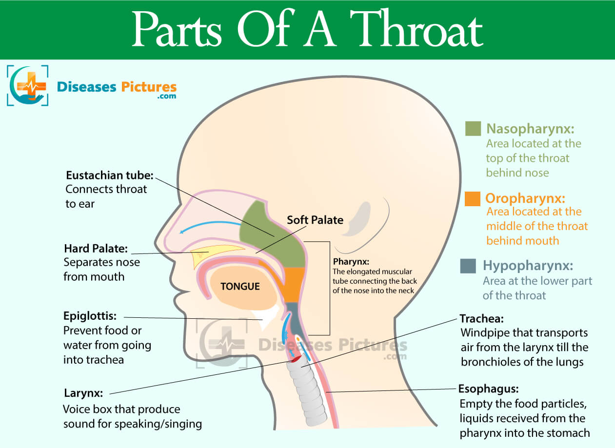
Throat Anatomy Throat Parts, Pictures, Functions HealthMD
Mouth. The mouth, or oral cavity, is the first part of the digestive tract.It is adapted to receive food by ingestion, break it into small particles by mastication, and mix it with saliva.The lips, cheeks, and palate form the boundaries. The oral cavity contains the teeth and tongue and receives the secretions from the salivary glands.. Lips and Cheeks. The lips and cheeks help hold food in.

Mouth Diagrams Printable 101 Diagrams
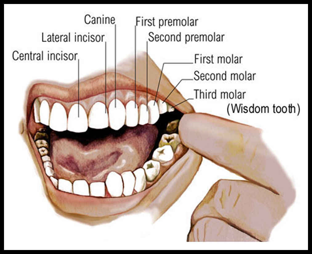
Mouth A molar tooth is located in the posterior (back) section of the mouth. It is found in most mammals that use their posterior teeth to grind food. Twelve molars are usually present in an.
Mouth diagram Healthiack
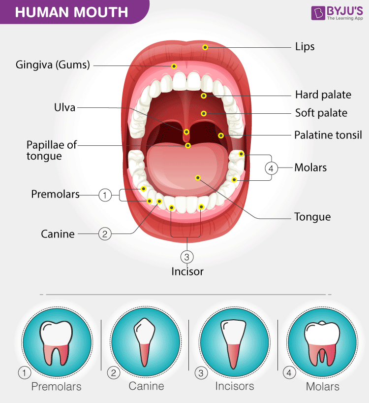
Lips Lips form the border of our mouth. Lips are a different color than the rest of our face because the skin around them is much thinner. Teeth and Gums The teeth are used to break up the foods that we eat. Teeth are made from enamel, the hardest substance found in our body.
The Anatomy Of The Mouth
When we say 'mouth' we mean the oral cavity; a space in the lower part of the head that functions as the entrance to the digestive system. The content of the oral cavity determines its function. It houses the structures necessary for mastication and speech, which include the teeth, the tongue and associated structures such as the salivary glands.
Mouth Teeth Diagram with Label Health Images Reference
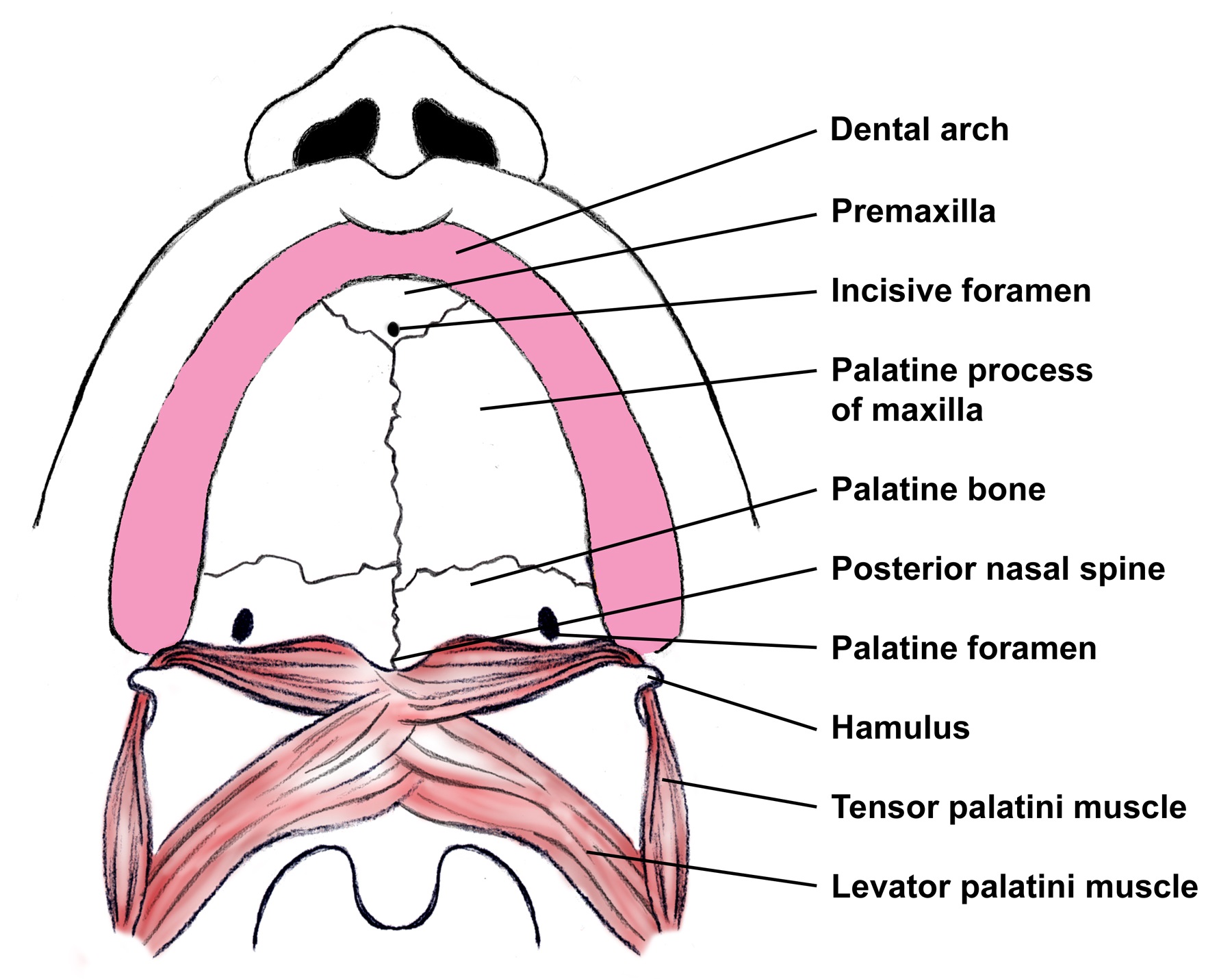
Anatomy of the Oral Cavity. Figure 1. Anterior view of the A external mouth and lips and B arterial supply to the lips. Figure 2. Inferior view of the maxilla. Figure 3. Cross section of a tooth. Figure 4. Lateral cross-section showing the A innervation of the lips B and teeth and gingiva.
Anatomy of the Mouth
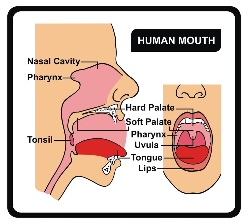
The pharynx (throat) is involved in both digestion and respiration. It receives food and air from the mouth, and air from the nasal cavities. When food enters the pharynx, involuntary muscle contractions close off the air passageways. Figure 6. The pharynx runs from the nostrils to the esophagus and the larynx.
Mouth Diagrams Printable 101 Diagrams
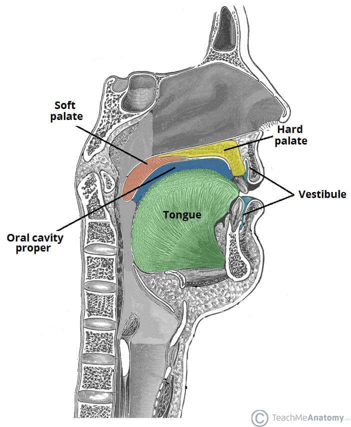
The oral cavity spans between the oral fissure (anteriorly - the opening between the lips), and the oropharyngeal isthmus (posteriorly - the opening of the oropharynx). It is divided into two parts by the upper and lower dental arches (formed by the teeth and their bony scaffolding).
Human Mouth Anatomy
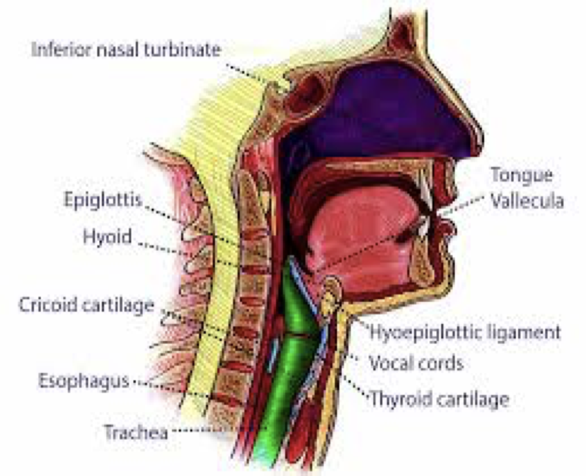
The tongue is a mobile, muscular organ that lies within the mouth and partly extends into the upper throat. The tongue's anatomy is complex; it involves interlacing muscles, nerves, and a blood supply. This article will explain the details of tongue anatomy and how each part contributes to its movements and to functions such as eating, taste.

AN3 08 Oral Cavity, Oropharynx, Swallowing StudyBlue
The main open area of the mouth, or oral cavity proper, runs from the gums and teeth to the fauces. When you are chewing, you do not find it difficult to breathe simultaneously. The next time you have food in your mouth, notice how the arched shape of the roof of your mouth allows you to handle both digestion and respiration at the same time.

Diagram of the Mouth 101 Diagrams
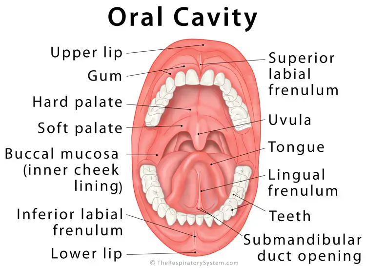
The oral cavity, or more commonly known as the mouth or buccal cavity, serves as the first portion of the digestive system. It consists of several different anatomically different aspects that work together effectively and efficiently to perform several functions. These aspects include the lips, tongue, palate, and teeth.
What is the Oral Cavity
What is the mouth? Your mouth is an oval-shaped opening that sits just below your nose. It starts at your lips and ends towards your tonsils. Your mouth is part of your digestive system and respiratory system. Other names for your mouth include oral cavity. Advertisement Cleveland Clinic is a non-profit academic medical center.

Printable Mouth Diagrams 101 Diagrams
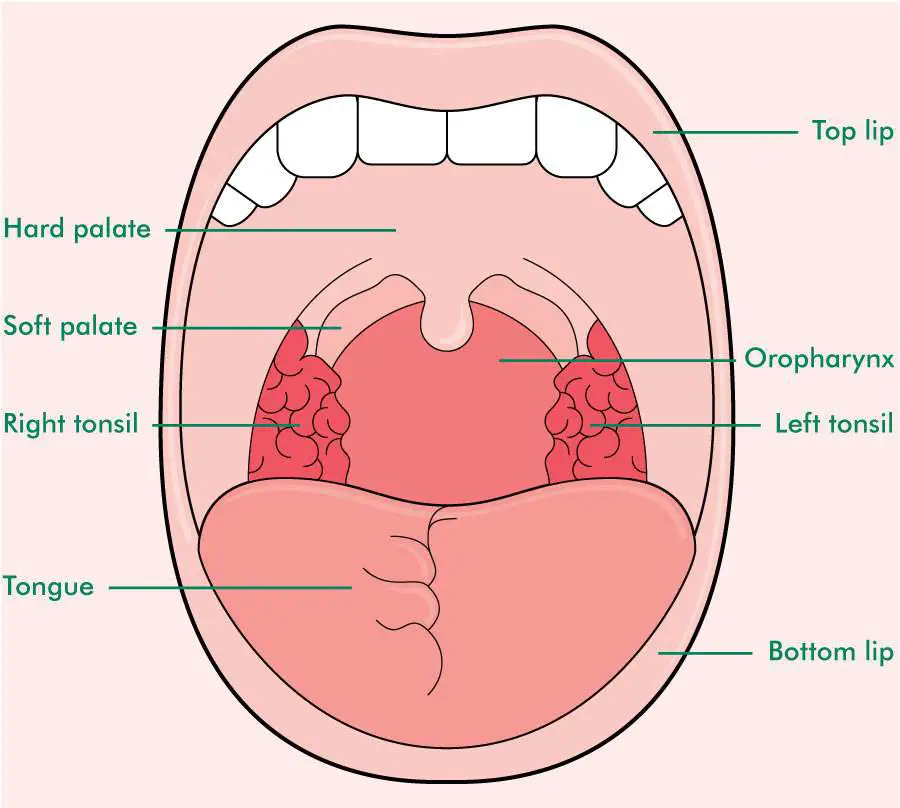
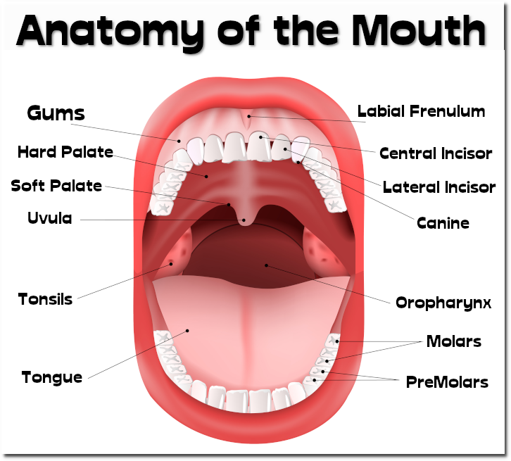
The boundaries of the oral cavity include the hard palate and soft palate that form the roof of your mouth, the tongue and the muscles below it, which make up the floor of the mouth and the inner surface of the lips in the front, the cheeks on the sides, and the uvula (the little "punching bag" shaped structure) at the end of your soft palate in.

The Mouth, Pharynx, and Esophagus Anatomy and Physiology II
Human mouth In human anatomy, the mouth is the first portion of the alimentary canal that receives food and produces saliva. [2] The oral mucosa is the mucous membrane epithelium lining the inside of the mouth. In addition to its primary role as the beginning of the digestive system, the mouth also plays a significant role in communication.

Oral cavity anatomy with educational labeled structure vector illustration
The mouth, also called the oral cavity, is the opening in the human skull that allows food, liquids, and air to enter the body. The oral cavity begins at the lips and ends at the throat. What are.
The Mouth and Buccal Cavity Anatomy of the Human Mouth
We have created 110 medical original illustrations of the mouth, the buccal cavity, the bones of the palate, the tongue, the salivary glands and the oral part of the pharynx with vessels and nerves.
The Oral Cavity Divisions Innervation TeachMeAnatomy
The mouth (oral cavity) consists of several components, including the teeth, gingiva (gums), tongue, palate, cheeks, lips and floor of the mouth. With the exception of the teeth, the mouth is lined by mucous membranes. The Teeth The teeth are held within the jaw bones and serve several important functions beyond allowing you to chew.
