Anatomy of the Spinal Cord TrialExhibits Inc.

Nervous and Spinal Cord Models
NERVOUS SYSTEM - nEURON & SPINAL CORD MODELS . Labeled Models. Leader-lined Models. Labeled Models. San Diego Mesa College 7250 Mesa College Drive San Diego, CA 92111-4998 Student Support San Diego Community College District San Diego City College San Diego Mesa College San Diego Miramar College

Anatomy of the Spinal Cord TrialExhibits Inc.
The spinal cord is a long, thin, tubular structure made up of nervous tissue that extends from the medulla oblongata in the brainstem to the lumbar region of the vertebral column (backbone) of vertebrate animals. The center of the spinal cord is hollow and contains a structure called central canal, which contains cerebrospinal fluid.

Spinal Cord Model Bing Images Spinal cord anatomy, Basic anatomy
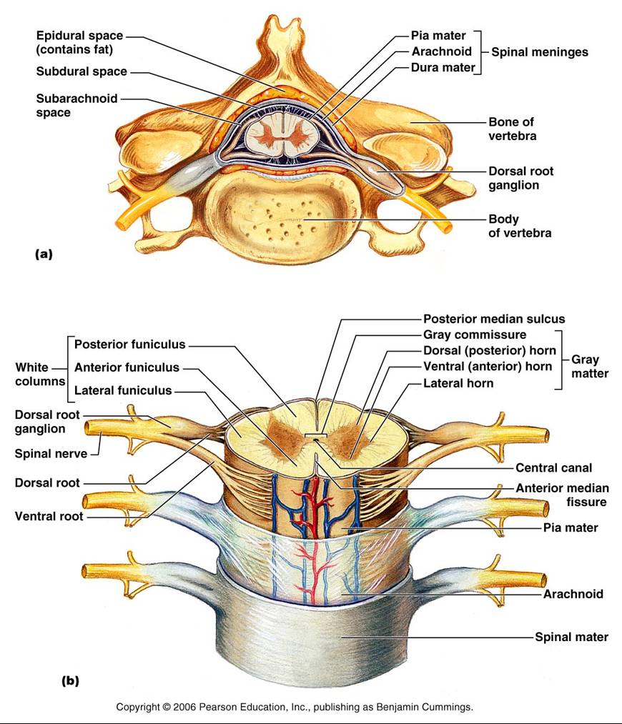
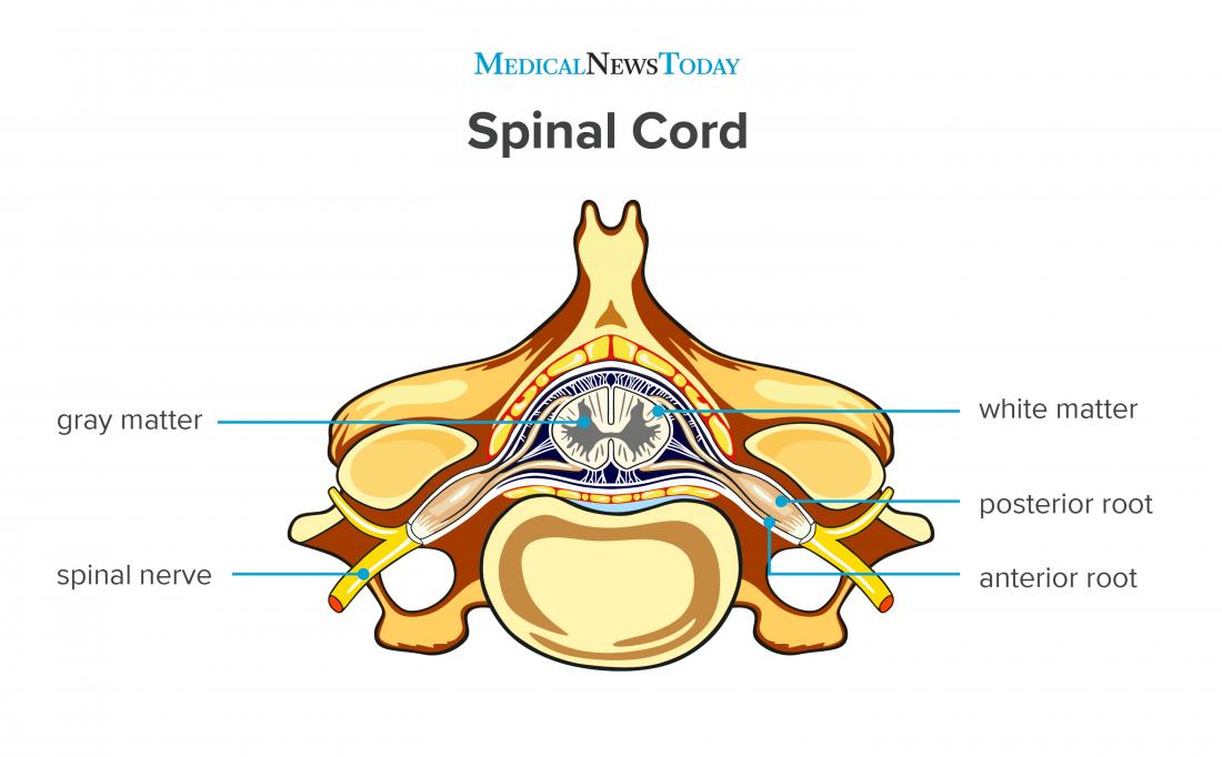
The spinal cord is a tubular bundle of nervous tissue and supporting cells that extends from the brainstem to the lumbar vertebrae.Together, the spinal cord and the brain form the central nervous system. In this article, we shall examine the macroscopic anatomy of the spinal cord - its structure, membranous coverings and blood supply.
BIOL 237 Class Notes The Spinal Cord and Spinal Nerves
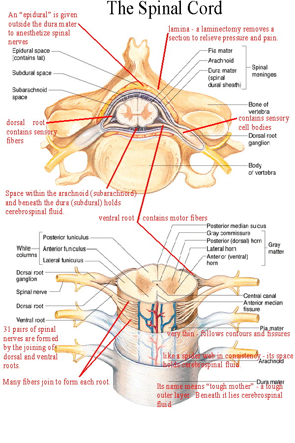
The spinal cord is part of the central nervous system (CNS), which extends caudally and is protected by the bony structures of the vertebral column. It is covered by the three membranes of the CNS, i.e., the dura mater, arachnoid and the innermost pia mater. In most adult mammals it occupies only the upper two-thirds of the vertebral canal as the growth of the bones composing the vertebral.

Spinal Cord Model Bing Images Nervous system anatomy, Human anatomy
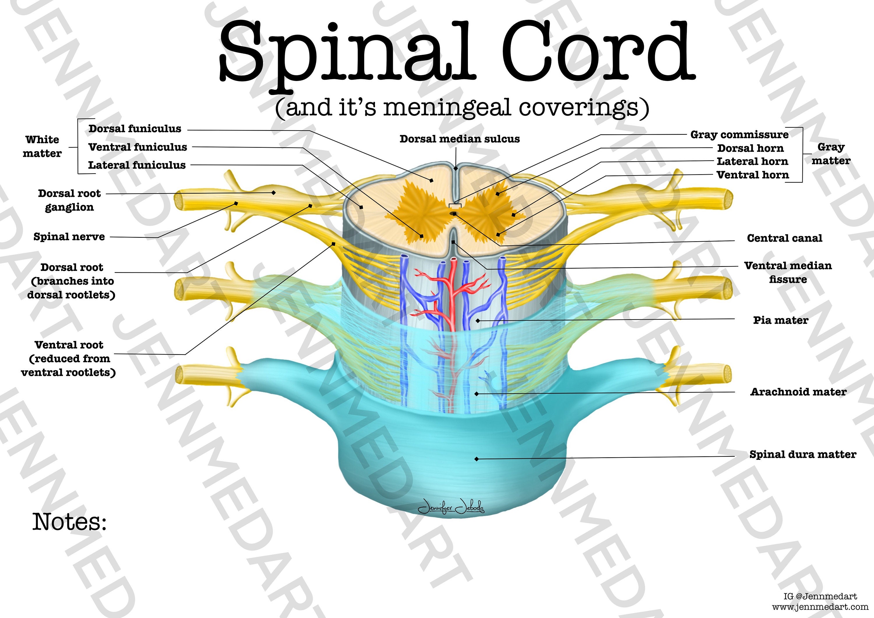
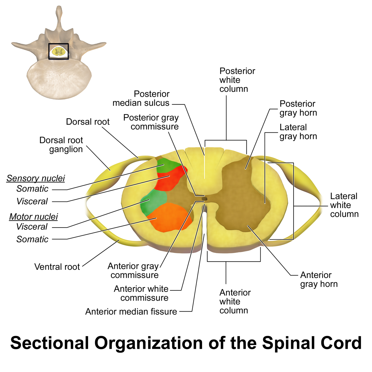
The spinal cord, along with the brain, makes up the central nervous system (CNS). It is a long tubular structure comprised of nervous tissue, extending from the cervical to the lumbar region of the vertebral column. Just like other parts of the CNS, the spinal cord is comprised of white and gray matter. Spinal cord gray matter is the central.
Ascending tracts of the spinal cord Anatomy Kenhub
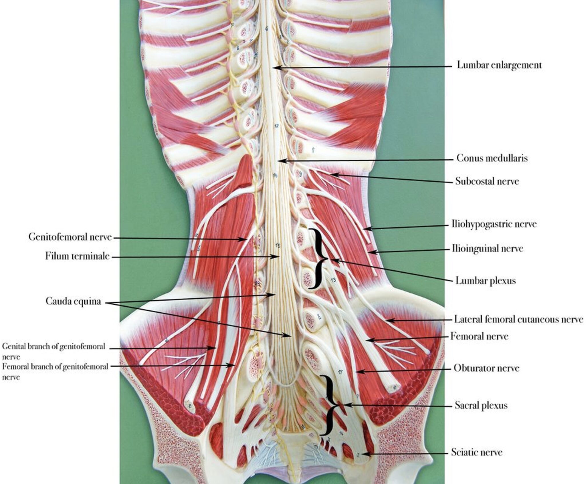
Figure \(\PageIndex{1}\): Gross Anatomy of the Spinal Cord. The spinal cord is divided into four regions: cervical, thoracic, lumbar and sacral. The sacral region has a tapered end called the conus medullaris. The bundle of axons inferior to the conus medullaris is the cauda equina. The cauda equina is anchored to the coccyx by the filum.
BY 411 Advanced Human Anatomy Blog Neuroanatomy Post 3 Development
Function What is the purpose of the spinal cord? Your spinal cord's main purpose is to carry nerve signals throughout your body. These nerve messages have three crucial functions. They: Control body movements and functions. Signals from your brain to other body parts control your movements.
Labeled Spinal Cord Model
The spinal cord is a continuation of the brainstem. It extends from the foramen magnum at the base of the skull to the L1/L2 vertebra where it terminates as the conus medullaris (medullary cone).

17 Best images about Anatomy Lab 2 on Pinterest Models, Human anatomy
Overview of spinal cord anatomy The spinal cord is a cylindrical mass of neural tissue extending from the caudal aspect of the medulla oblongata of the brainstem to the level of the first lumbar vertebra (L1).While the length of the spinal cord varies from one individual to another, it is usually longer in males (approximately 45 cm) than it is in females (approximately 42 cm).

Using a Spinal Cord Anatomical Model in the Courtroom
Looking for Spinal Model? We have almost everything on eBay. But did you check eBay? Check Out Spinal Model on eBay.

Spinal Cord Anatomy Model template
This atlas of human anatomy describes the spinal cord through 18 anatomical diagrams with 270 anatomical structures labeled. It was designed particularly for physiotherapists, osteopaths, rheumatologists, neurosurgeons, orthopedic surgeons and general practitioners, especially for the study and understanding of medullary diseases.
Spinal Cord Anatomy Worksheet Single FILLED Digital Download Human
The spinal cord is a part of the central nervous system (CNS) along with the brain. It is located within the vertebral canal of the spine. In the cranial direction, the spinal cord is continuous with the medulla oblongata of the brainstem. In the caudal direction, it terminates as the medullary cone (conus medullaris).
Spinal Cord Summary Neuroanatomy Geeky Medics
Explore Our Excellent Selection Of Anatomical Spine Models. Shop & Save Today! Our Most Popular Spine Model For Patient & Student Education Is Also Our Most Affordable
Spinal cord Anatomy, functions, and injuries
The vertebra provides several crucial functions to the body. First, it acts as a structural component of the spine, bearing body weight, anchoring muscles and the spinal cord, and forming joints with other vertebrae and ribs that allow the torso and neck to move. Second, the vertebra protects the delicate tissues of the spinal cord by.
Labeled Spinal Cord Model
In these topics. Quick Facts: Injuries of the Spine and Spinal Cord Quick Facts: Overview of Spinal Cord Disorders Injuries of the Spinal Cord and Vertebrae Cervical Spinal Stenosis Lumbar Spinal Stenosis Neck Pain Sciatica Herniated Disk Low Back Pain Overview of Spinal Cord Disorders.

Nervous and Spinal Cord Models
The spinal cord is a mass of nervous tissue that extends inferiorly from the brain stem through the vertebral canal of the cervical and thoracic regions, ending around the T12 or L1 vertebra.mycontentbreak It is a long tube about 18 inches (45 cm) in length and around half an inch (1 cm) in diameter at its widest point.
