How the Human Eye Works (Structure and Function)

Eye Anatomy
Structure and Functions of Human Eye with labelled Diagram Biology Biology Article Structure Of Eye Structure of the Eye The eye is one of the sensory organs of the body. In this article, we shall explore the anatomy of the eye The structure of the eye is an important topic to understand as it one of the important sensory organs in the human body.
eye anatomy Optometrist in Petaling Jaya Optical Shop Promotion Malaya Optical
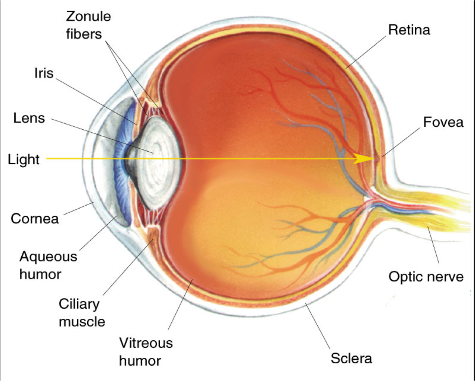
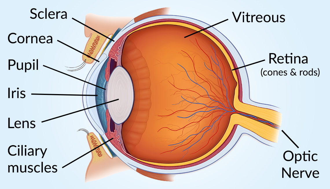
The iris (colored part) of the eye functions like the diaphragm of a camera, controlling the amount of light reaching the retina by automatically adjusting the size of the pupil (aperture). The eye's crystalline lens is located directly behind the pupil and further focuses light rays.

Blind Spot Eye Anatomy ANATOMY
Diabetes Healthy ANATOMY and Eyes OF THE AND ITS FUNCTION Toolkit Parts of the Eye Vision is wonderful, but you could lose To understand it if you eye have problems, diabetes. it is helpful to know the different parts of the eye. Please refer to the back of this handout for descriptions of their functions. The main parts of the eye— Optic 3
Eye Diagram Cliparts.co
Eyelid anatomy Lacrimal gland Eye muscles Eyeball Outer layer Middle layer Inner layer Blood supply of the eye Nerves of the eye Sources + Show all Bones of the orbit The bony orbit is made out of seven bones, which include the maxilla, zygomatic bone, frontal bone, ethmoid bone, lacrimal bone, sphenoid bone and palatine bone.

Anatomy of the Eye Human eye diagram, Eye anatomy diagram, Eye anatomy
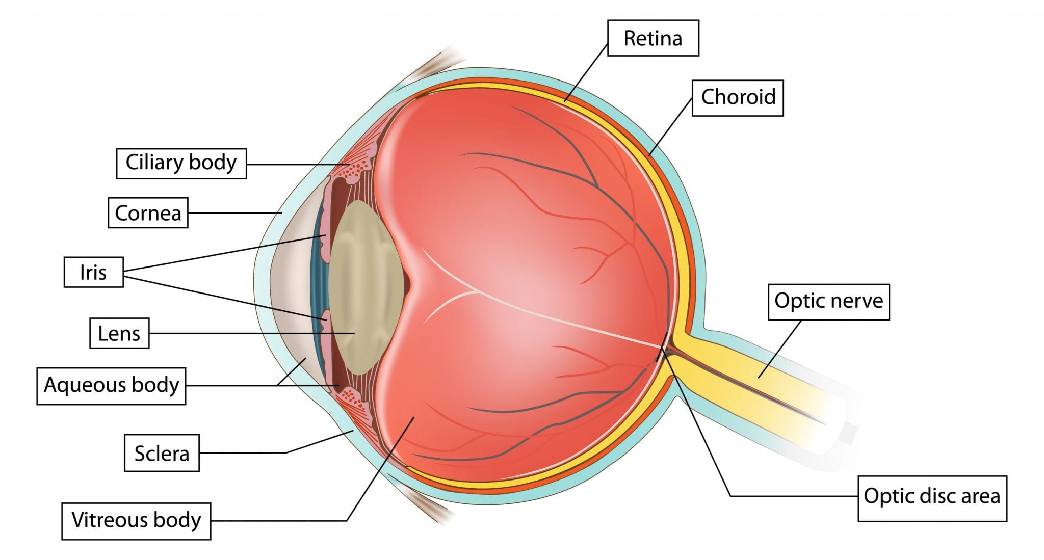
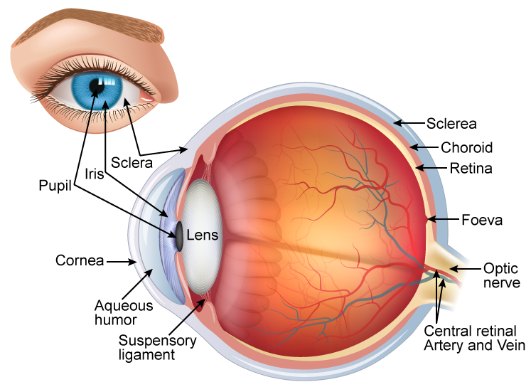
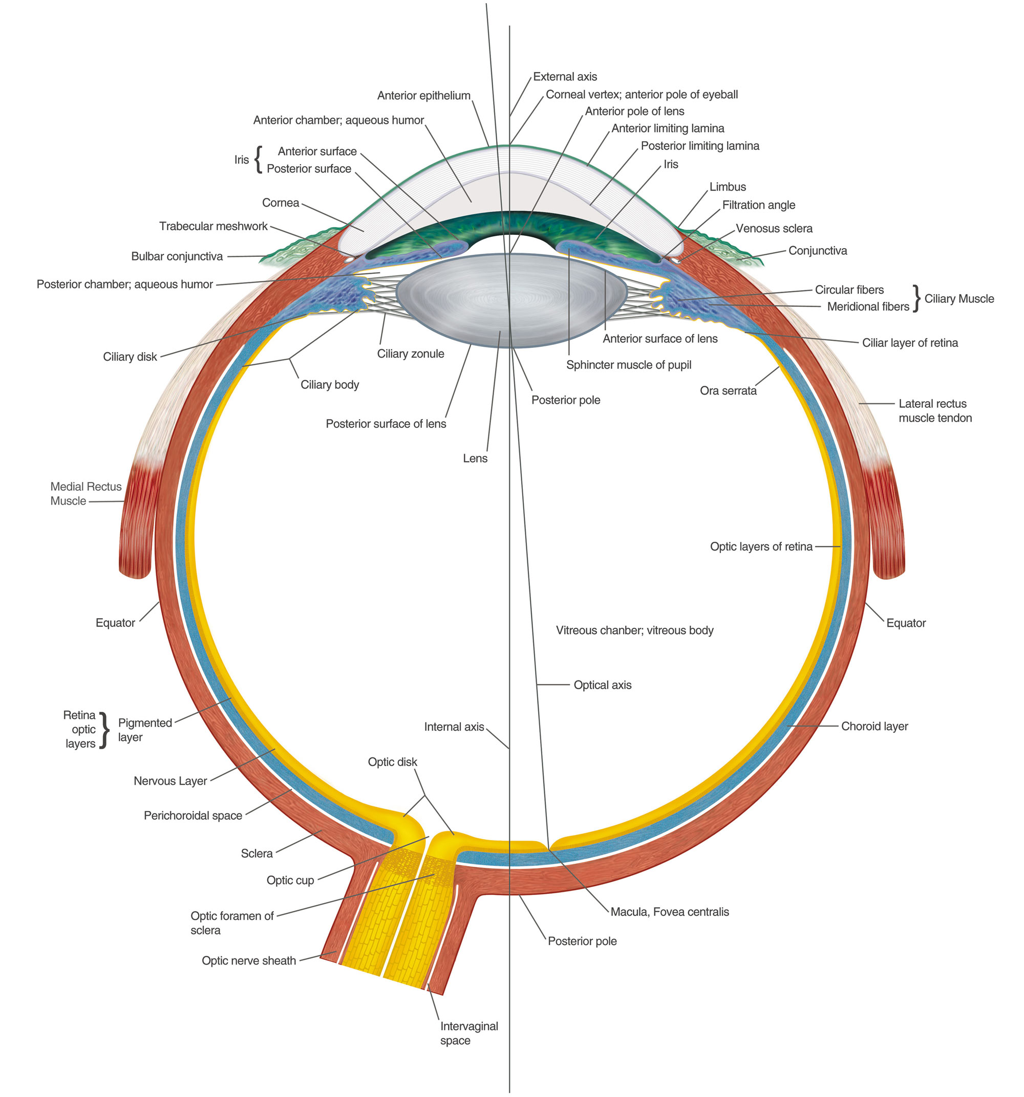
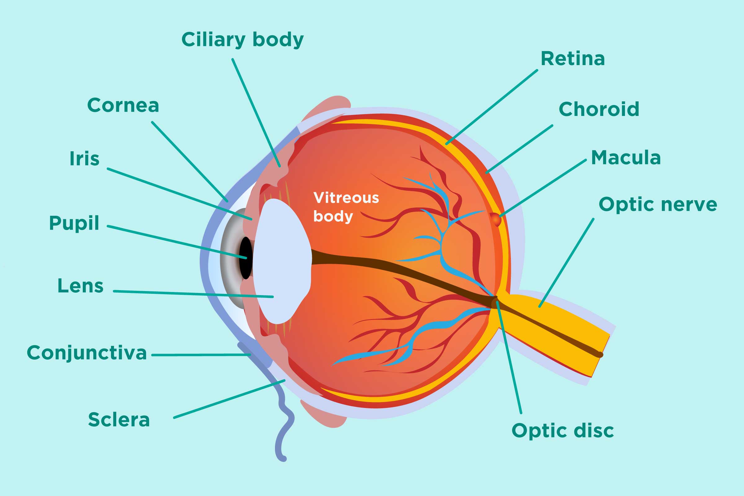
A brief description of the eye along with a well-labelled diagram is given below for reference. Well-Labelled Diagram of Eye The anterior chamber of the eye is the space between the cornea and the iris and is filled with a lubricating fluid, aqueous humour. The vascular layer of the eye, known as the choroid contains the connective tissue.

OUR EYES WORK LIKE CAMERA’S! Discovery Eye Foundation
Diagram of the Eye Posted in Eye Health, Uncategorized | August 5, 2018 Even though the eye is small, only about 1 inch in diameter, it serves a very important function - the sense of sight.

Diagram showing the different parts of the eye Parts of the eye, Eye health, Free homeschool
Download. English: Parts of the Eye (PDF 603.5 KB) Spanish: Las partes del ojo (PDF 897.7 KB) Check out this fact sheet to see a labeled diagram of the eye and learn about the different parts of the eye.
draw a neat and labelled diagram of structure of the human eye slwbyx77 Science
1. Conjunctiva The conjunctiva is the membrane covering the sclera (white portion of your eye). The conjunctiva also covers the interior of your eyelids. Conjunctivitis, often known as pink eye, occurs when this thin membrane becomes inflamed or swollen. Other eye disorders that affect the conjunctiva include:
ARCHIVE FileAnatomy of the eye.jpg Comparative Physiology of Vision
The iris controls widening and narrowing (dilation and constriction) of the pupil. Cornea: the transparent circular part of the front of the eyeball. It refracts the light entering the eye onto the lens, which then focuses it onto the retina. The cornea contains no blood vessels and is extremely sensitive to pain.

Can We Grow New Eyes?
The retina is the innermost layer lining the back of the eyeball and the light-sensitive part of the eye. The retina contains photoreceptors that detect light. These photoreceptors are known as cones and rods. Cones enable us to detect colours, while rods help us to see in poor light. The retina contains nerve cells that transmit signals from.

Human eye Extraocular Muscles Britannica
The structures and functions of the eyes are complex. Each eye constantly adjusts the amount of light it lets in, focuses on objects near and far, and produces continuous images that are instantly transmitted to the brain. The orbit is the bony cavity that contains the eyeball, muscles, nerves, and blood vessels, as well as the structures that.
Vision and Eye Diagram How We See
Labeled diagram of the eye Unlabeled diagram of the eye Eye anatomy quizzes Sources + Show all How to learn the parts of the eye Found within two cavities in the skull known as the orbits, the eyes are surrounded by several supporting structures including muscles, vessels, and nerves.
/GettyImages-695204442-b9320f82932c49bcac765167b95f4af6.jpg)
Structure and Function of the Human Eye
Apr. 29, 2023 To understand the diseases and conditions that can affect the eye, it helps to understand basic eye anatomy. Here is a tour of the eye starting from the outside, going in through the front and working to the back. Eye Anatomy: Parts of the Eye Outside the Eyeball The eye sits in a protective bony socket called the orbit.
Inflammatory Arthritis and Eye Health Prevention, Symptoms, Treatment
Labelling the eye. Use this interactive to label different parts of the human eye. Drag and drop the text labels onto the boxes next to the diagram. Selecting or hovering over a box will highlight each area in the diagram. The human eye has several structures that enable entering light energy to be converted to electrochemical energy.

Human Eye Anatomy Parts of the Eye and Structure of the Human Eye
6 min read Your eye is a slightly asymmetrical globe, about an inch in diameter. The front part (what you see in the mirror) includes: Iris: the colored part Cornea: a clear dome over the iris.

File1413 Structure of the Eye.jpg Wikimedia Commons
There are six extraocular muscles that attach to the outside of the eye from the bone in the eye's socket. These muscles work to rotate the eye, and move the eye up, down, and from side to side. When they work together, these muscles can move the eye in any direction. Medial Rectus (MR) - Moves the eye inward, towards the nose.
